Epigenetics Meets CRISPR/Cas to Fight Cancer
Received Date: January 23, 2019 Accepted Date: February 28, 2020 Published Date: March 03, 2020
doi: 10.17303/jcrto.2020.8.202
Citation: Simeon Santourlidis (2020) Epigenetics Meets CRISPR/Cas to Fight Cancer. J Cancer Res Therap Oncol 8: 1-5.
Abstract
Epigenetic changes are fundamental for cancer. To our knowledge, these are the only ones that include alterations that are consistently present in many cancer entities at once. Hence they may provide targets for general cancer therapy. Now, fortunately, the exciting in-vivo genome targeting CRISPR/Cas technology has been developed and one new perspective has arisen to take advantage of these common epigenetic targets to fight cancer.
CRISPR/Cas differential substrate and chromatin specificity
Clustered regularly interspaced short palindromic repeats ((CRISPR)/CRISPR-associated (Cas)) technology holds the potential to decisively impact almost all disciplines in current biological and medical research. For that reason, it is currently discussed how it may contribute to new cancer gene therapy strategies. Here, I wish to suggest one approach to apply this new CRISPR/Cas technology to target cancer cells based on their aberrant chromatin structure. I will discuss this approach primarily for urological cancers, my subject of research, but in addition, it should be promising to be applied in a wide range of cancer entities.
CRISPR/Cas systems evolved as widespread adaptive immunity systems that protect bacteria and archaea against phages and plasmids [1] by directing sequence-specific Cas9 endonuclease mediated double-strand DNA cleavage (DSB) to the intruder´s DNA and hence destruct its genetic information [2]. In its native species, this system has evolutionary never encountered as a substrate DNA which is organized in complex, high-order structured chromatin, which serves in mammalian cells to control accessibility, transcription and epigenetic functionality. One would therefore intuitively expect that Cas9 cleavage of eukaryotic DNA, especially if organized as heterochromatin, would be less efficient than that of its original substrate. Indeed, evidence in multiple reports indicates that Cas9 has a high preference for binding to more easily accessible chromatin regions [3- 5] consistent with the observation that active sg RNAs map in the areas of open chromatin [6]. Furthermore, active transcription can directly stimulate DNA cleavage by CRISPR/Cas. This has been shown on allele-specific euchromatic and heterochromatic, respectively, states of the p16INK4a locus CpG-island [7] and for imprinted alleles where the repressed heterochromatic allele accumulated Cas9 mutations slower than the active one. Thus tightly packed heterochromatin nucleosomes negatively affect Cas9 binding and functioning [8].
Adjustment of CRISPR/Cas methodological parameters to trigger efficiency
Brief exposure and low Cas9 expression confer further differences in efficiency [9], indicating that differential efficiency can be further adjusted by sophisticatedly selected methodological parameters. Obviously, the system offers a wide spectrum of possible adjustments in order to optimize efficiency and target specificity. For instance, the ratio of on- to off-target cleavage is improved by the reduction of the concentrations of sgRNA and Cas9 nuclease expressed in the cell [10]. The specificity of the nuclease is complex and target site-dependent. For instance, single and double mismatches are often well tolerated even when one or more mismatches occur in the 3‟ half of the sgRNA targeting sequence but not all mismatches in the 5‟ half of the sgRNA/ DNA interface are necessarily well tolerated [10]. Furthermore, a high GC-content, as e.g. exists in the Long Interspersed Nuclear Element 1 (LINE-1) promoter region and in CpG islands, has been shown to stabilize RNA: DNA hybrids and is therefore expected to make sgRNA/genomic DNA hybrids more stable and more tolerant to mismatches, i.e. to enhance on-target specificity. On the other hand two or more intentionally interspaced mismatches dramatically reduce Cas9 cleavage [10,11] Finally it is mentioned that the rapidly evolving Cas9 research field is steadily providing newly discovered or recombinantly modified Cas variants and Cas9 homologs with a plethora of new and adjusted, respectively, features which e.g. reduce non-specific interactions and contribute to an improved efficiency and specificity [12].
Characteristic configuration of cancer chromatin and the example of LINE-1
Cancer cell-specific reorganization of chromatin configuration leads to microscopically visible, profound alterations in chromatin structure which include dense “hyperchromatic” chromatin. This is a hallmark of cancer known since 1914, first described by Teodor Boveri [13] and one criteria to distinguish cancer cells in routine pathology. For instance, it has been exemplary exploited worldwide, for over 65 years clinically applied, ‟Pap test”, a central pillar of screening for cervical cancer which has probably saved millions of lives, constituting a true public health success [14]. An outstanding and motivating achievement by George Papanicolaou in the 1930s [15].
One basic assumption underlying my considerations outlined below is that within their huge “chromatinome”, the majority of cells of one cancer entity share common chromatin signatures, which contain consistently euchromatic regions, which are heterochromatic in the corresponding healthy cells of origin. These would constitute primary targets for intentional, differentially effective operating Cas9 endonucleolytic disruption. Comparative global chromatin analyses between a significant number of primary tumors and the appropriate cells of origin should reveal such cancer-specific, consistent euchromatic signatures and I hypothesize that this would be possible for presumably many cancer entities.
One issue I have been working on in the last two decades, namely the epigenetic deregulation of Long Interspersed Nuclear Elements 1 (LINE-1) retroelements in cancer, provides a first paradigm and an exciting opportunity in this respect.
In all healthy somatic cells, LINE-1 retroelements are densely methylated at their CpG dense promoter regions, tightly packaged into inactive chromatin and poorly accessible for transcription [16]. This state is established in healthy differentiated somatic cells during ontogenesis by coordinately acting epigenetic mechanisms to ensure repression of these dispersed genetic elements which otherwise could become activated and mobile, threatening the integrity of the genome. LINE-1 elements of healthy differentiated somatic cells typically bear histone H3 methylated at Lys9 (H3K9me2/me3) as a mark of repressed chromatin [17] are enriched in heterochromatic regions of the genome as shown by bioinformatics data [18] and experimentally by FISH [19,20]. They are associated with H2A.Z, a repressive histone variant and HP1α, a canonical heterochromatin protein repressing transcription [21] while showing an anti-correlation with euchromatic marks [22,23]. Noteworthy, the γH2AX signal which marks DNA double-strand breaks is weaker in heterochromatin and LINE-1 elements presented a lower γH2AX signal after irradiation with 2 Gy X-rays [22]. This observation indicates that LINE-1s are indeed situated in heterochromatic regions that are less susceptible to the induction of DNA double- strand breaks than euchromatic regions.
In addition, recent evidence demonstrates that the silencing of LINE-1 elements decreases chromatin accessibility, whereas activation prevents chromatin compaction and suggests that LINE-1 functions primarily at the chromatin level and acts as a global chromatin accessibility regulator [24].
This repressed state is alleviated in many cancers. A loss of DNA methylation in the CpG rich LINE-1 promoter sequences is documented to occur in a broad variety of cancers, e.g. colon, lung, prostate and breast cancer, hepatocellular and gastric carcinoma, chronic lymphocytic leukemia (CLL) and this loss contribute to “genome-wide” or “global” DNA hypomethylation [25,27]. Loss of methylation at LINE-1 promoters is thought to favor a gain of transcriptional competence. It is estimated that about 3000 full-length LINE-1s exist in a human genome, of which about 100 contain intact open reading frames [26], which could become expressed if they became more euchromatic and accessible. DNA hypomethylation of LINE-1 retrotransposons is, in particular, an early and very frequent epigenetic event during urothelial carcinogenesis [25], occurring in over 90% of all cases [28,29]. There is accumulating evidence that this is associated with increased levels of full-length LINE-1 transcripts [30]. Of note, LINE-1 hypomethylation is associated with cancer progression, becoming more pronounced in high-stage and high-grade cancer [31,32].
The example of prostate and Epi CRISPR/ Cas Cancer Therapy (ECCT)
In contrast to bladder cancer, global LINE-1 hypomethylation is a late event in prostate carcinogenesis and is clearly associated with tumor progression [31,28,33]. Here hypomethylation increases with tumor grade and stage and particularly pronounced hypomethylation is seen in lymph node-positive tumors [31]. Analyses on mortality from prostate cancer stratified for the Gleason score revealed that LINE-1 hypomethylation was associated with mortality in patients with higher Gleason scores of at least 8 [33]. These data suggest that increased mortality from prostate cancer is associated with lower levels of LINE-1 methylation in the tumor tissue.
In conclusion, I wish to suggest here that CRISPR/Cas targeting of hypomethylated LINE-1 elements in cancer could lead to a selective disruption of cancer DNA and subsequently cause cancer cell death while sparing healthy somatic cells. It could be helpful that these repetitive DNA elements are scattered in large numbers over the whole genome, presenting multiple targets, so that it might be possible to overcome residual cancer cell DNA repair mechanisms by simply adjusting the above discussed methodological parameters. Among DNA damages, double-strand breaks (DSBs) are one of the most harmful, considered to be lethal lesions to a cell [34]. A hierarchical signaling pathway is orchestrated by various proteins that sense DNA damages, transduces the signals to the effectors, and determines the cell fate [35]. When DNA damages are too severe and unrepairable, cells are forced to undergo programmed cell death [35]. Noteworthy, the expression of DSB repair genes is disturbed in various cancers, which has been exploited by using genotoxic agents to generate DNA double-strand breaks as an intermediate in radio- and chemotherapy strategies [34].
Notably, cancer cell-specific facultative euchromatic chromatin signatures extend to sequences beyond LINE-1, which may serve as further potential CRISPR/Cas targets in addition to LINE-1.
As with any genetic approach, delivery is expected to present a major hurdle to the application of the ECCT. In this respect, superficial bladder cancer could be used to test this approach based on accessibility through the transurethral application, ease of administration, limited systemic dissemination, as routinely used in the application of BCG immunotherapy, but also experimentally for virus-based gene and immunotherapy [36]. Moreover, healthy uroepithelial cells of the bladder are covered by surface glycans, specialized lipid molecules, and uroplakins at their apical surface which ensures the low permeability of the urothelium and provides a barrier function which protects it from extrinsic and toxic components of urine [37]. Furthermore, in contrast to the rapid cell division of urothelial tumors, the healthy uroepithelial cells of the bladder have a low rate of the division-leading to a turnover rate of the uroepithelium of ∼3–6 mo [38]. Both characteristics would additionally support the differential response of uroepithelial cancer cells compared to uroepithelial cells in an in vivo application of a CRISPR/Cas euchromatic LINE-1 targeting therapeutic approach. Noteworthy, CRISPR/Cas9 cancer cell specificity and its therapeutic effects could be further enhanced by using appropriate delivery vehicles, e.g. vaccinia or retroviruses that infect only dividing cells.
Acknowledgments
I dedicate this article in gratefulness to my mentor Prof. Wolfgang A. Schulz.
- Deltcheva E, et al. (2011) CRISPR RNA maturation by trans-encoded small RNA and host factor RNase III. Nature 471: 602-607.
- Jinek M, et al. (2012) A programmable dual-RNA-guided DNA endonuclease in adaptive bacterial immunity. Science 337: 816-821.
- Kuscu C, et al. (2014) Genome-wide analysis reveals characteristics of off-target sites bound by the Cas9 endonuclease. Nat. Biotechnol 32: 677-683.
- O'Geen H, et al. (2015) A genome-wide analysis of Cas9 binding specificity using ChIP-seq and targeted sequence capture. Nucleic Acids Res 43: 3389-3404.
- Wu X, et al. (2014) Genome-wide binding of the CRISPR endonuclease Cas9 in mammalian cells. Nat. Biotechnol 32: 670-676.
- Radzisheuskaya A, et al. (2016) Optimizing the sgRNA position markedly improves the efficiency of CRISPR/dCas9- mediated transcriptional repression. Nucleic Acids Res. 44:104.
- Fujita T, et al. (2016) Allele-specific locus binding and genome editing by CRISPR at the p16INK4a locus. Sci. Rep 28: 6.
- Verkuijl SA and Rots MG (2019) The influence of eukaryotic chromatin state on CRISPR- Cas9 editing efficiencies. Curr. Opin. Biotechnol 55: 68-73.
- Kallimasioti-Pazi EM, et al. (2018) Heterochromatin delays CRISPR-Cas9 mutagenesis but does not influence the outcome of mutagenic DNA repair. PLoS Biol 16:12.
- Fu Y, et al. (2013) High-frequency off-target mutagenesis induced by CRISPR-Cas nucleases in human cells. Nat. Biotechnol 9: 822-826.
- Hsu PD, et al. (2013) DNA targeting specificity of RNAguided Cas9 nucleases. Nat. Biotechnol 9: 827-832.
- Nakade S, et al. (2017) Cas9, Cpf1 and C2c1/2/3-What's next? Bioengineered 4: 265-273.
- Dietel M (2014) Boveri at 100: the life and times of Theodor Boveri. J. Pathol 234: 135-137.
- Bettigole C (2013) The thousand-dollar Pap smear. N. Engl. J. Med 369: 1486-1487.
- Papanicolaou GN (1942) A NEW PROCEDURE FOR STAINING VAGINAL SMEARS. Science 95: 438-439.
- Schulz WA (2006) L1 Retrotransposons in Human Cancers J. Biomed. Biotechnol 5.
- Goodier JL (2016) Restricting retrotransposons: a review. Mob. DNA 7: 16.
- Waterston RH, et al. (2002) On the sequencing of the human genome. Proc. Natl. Acad. Sci. U S A 99: 3712-3716.
- Boyle AL, et al. (1990) Differential distribution of long and short interspersed element sequences in the mouse genome: chromosome karyotyping by fluorescence in situ hybridization. Proc. Natl. Acad. Sci. U.S.A 87: 7757-7761.
- Solovei I, et al. (2009) Nuclear architecture of rod photoreceptor cells adapts to vision in mammalian evolution. Cell 137: 356-368.
- Rangasamy D (2013) Distinctive patterns of epigenetic marks are associated with promoter regions of mouse LINE-1 and LTR retrotransposons. Mob DNA 4: 27.
- Natale F, Scholl A, Rapp A, Yu W, Rausch C, et al. (2018) DNA replication and repair kinetics of Alu, LINE-1 and satellite III genomic repetitive elements. Epigenetics Chromati 11: 61.
- Bulut-Karslioglu A, et al. (2014) Suv39h-dependent H3K9me3 marks intact retrotransposons and silences LINE elements in mouse embryonic stem cells. Moll Cell 55: 277-290.
- Jachowicz JW, et al. (2017) LINE-1 activation after fertilization regulates global chromatin accessibility in the early mouse embryo. Nat. Genet 49: 1502-1510.
- Schulz WA, et al. (2006) Methylation of endogenous human retroelements in health and disease. Curr Top Microbiol Immunol 310: 211-250.
- Goodier JL, Kazazian HH Jr, (2008) Retrotransposons revisited: the restraint and rehabilitation of parasites. Cell 3: 23- 35.
- Wilson AS, et al. (2007) DNA hypomethylation and human diseases. Biochim. Biophys. Acta. 1775: 138-162.
- Schulz WA (1998) DNA methylation in urological malignancies (review). Int. J.Oncol 13: 151- 167.
- Neuhausen A, et al. (2006) DNA methylation alterations in urothelial carcinoma. Cancer Biol. Ther 5: 993-1001.
- Kreimer U, et al. (2013) HERV-K and LINE-1 DNA Methylation and Reexpression in Urothelial Carcinoma. Front Oncol 263.
- Santourlidis S, et al. (1999) High frequency of alterations in DNA methylation in adenocarcinoma of the prostate. Prostate 39: 166-174.
- Florl AR, Lower R, Schmitz-Drager BJ, Schulz WA, et al. (1999) DNA methylation and expression of LINE-1 and HERVK provirus sequences in urothelial and renal cell carcinomas. Br. J. Cancer 80: 1312-1321.
- Fiano, et al. (2017) LINE-1 methylation status in prostate cancer and non-neoplastic tissue adjacent to tumor in association with mortality. Epigenetics 12: 11-18.
- Srivastava M, and Raghavan SC (2015) DNA doublestrand break repair inhibitors as cancer therapeutics. Chem. Biol 22: 17-19.
- Karimian A, et al. (2019) CRISPR/Cas9 technology as a potent molecular tool for gene therapy. J. Cell. Physiol 234: 12267-12277.
- Burke J (2010) Virus therapy for bladder cancer. Cytokine Growth Factor Rev. 21: 99- 102.
- Wan Q, et al. (2018) Urothelium with barrier function differentiated from human urine-derived stem cells for potential use in urinary tract reconstruction. Stem. Cell. Res. Ther 9: 304.
- Khandelwal P, et al. (2009) Cell biology and physiology of the uroepithelium. Am. J. Physiol. Renal. Physiol 297: 1477-1501.
FIGURE 1
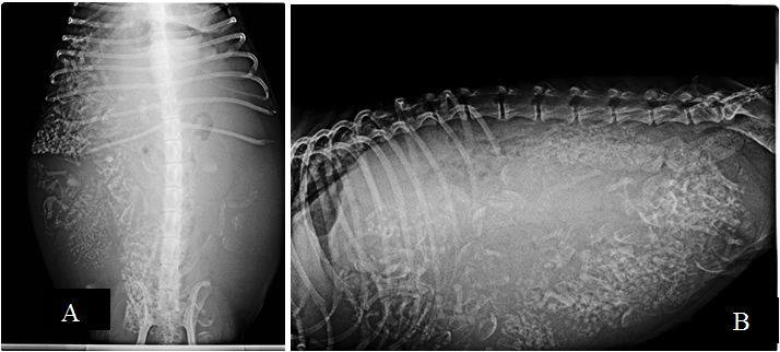
Figure 1: (A) Pointer bitch�s ventrodorsal abdominal radiograph that showing debris of fetuses� bones; (B) Golden retriever bitch�s laterolateral abdominal radiograph that showing debris of fetuses� bones
FIGURE 2
Figure 1: (A) Pointer bitch�s ventrodorsal abdominal radiograph that showing debris of fetuses� bones; (B) Golden retriever bitch�s laterolateral abdominal radiograph that showing debris of fetuses� bones
FIGURE 3
Figure 1: (A) Pointer bitch�s ventrodorsal abdominal radiograph that showing debris of fetuses� bones; (B) Golden retriever bitch�s laterolateral abdominal radiograph that showing debris of fetuses� bones
Tables at a glance
Figures at a glance