Ab Initio Analysis of Keratin (C6H12N2O4S2): A density functional approach
Received Date: January 26, 2023 Accepted Date: February 26, 2023 Published Date: February 28, 2023
doi: 10.17303/aicb.2023.1.102
Citation: Nirbhay Kumar Singh (2023) Ab Initio Analysis of Keratin (C6H12N2O4S2): A density functional approach. Ann Immunol Cell Biol 1: 1-10
Abstract
Keratin is a protein constituting high-sulphur content and form the bulk of epidermal appendages such as hair, nails, claws, turtle scats, horns, whale baleen, beaks, and feathers, it also help to regulate the size of cells, allow cells to move, grow, and divide heal wounds. It is also a building block of the human body and help in make up of the tissues in the skin, hair, and nails. Keratin wastes mostly produced from the poultry farms, slaughterhouses, and leather industries. It is also dumped, buried, used for landfilling, or incinerated, all these actions increase the threats of environmental hazards, pollution, negatively influence the public health, and increase greenhouse gases concentration.
Present paper is SIESTA based study on structural and physical properties of keratin by density function theory (DFT). Here we use molecular electrostatic potential (MEP) surface, Charge density analysis and partial density of state (PDOS) and density of state types (DOS) analysis were performed to analysed contribution of total total energy states, and each atomic orbitals in molecules. Bond length, bond angle, molecular surface were analysed by xcrysden and Jmol programme.
Keywords: Molecular Electrostatics Potential Surface (MEP). Projected Density of State (PDOS), Density of States (DOS), and Density function theory (DFT).
Introduction
Keratins are the main constituents of hair, nails, and hooves of mammals. It produce within cells and regenerative layer of the skin. It is a protective protein, less prone to scratching or tearing than other types of cells produces by our body. Keratin can be derived from the feathers, horns, and wool of different animals and used as an ingredient in hair cosmetics. Since it is the structural building block of our hair, therefore it is believe that keratin supplements, products, and treatments can help strengthen our hair and make it look healthier [1.2].
Smoothness of hair on nature of thickness and types of keratin treatment. Hair cuticle of hair cell theoretically absorb the keratin, due to which hair looks full and glossy. It can also make curly hair less frizzy, easier to style, and straighter in appearance.
Since keratin is a protein, it is important to eat protein-rich foods i.e. Eggs, onion, salmon, sweet potato which produce keratin. Certain nutrients like sweet potatoes are high in vitamin A, sunflower seeds, mango, garlic, kale etc. help the body produce keratin and may help improve the health of the skin, hair, nails, and other tissues [3,4].
Now a days DFT has become quite popular among researchers working in geoscience, biology, engineering and material science [5] here we use density function theory to analyse physical and chemical properties of Keratin. It is a successful theory and computationally less intensive then other method to calculate the electronic structure of atoms, molecules, and solids. Its goal is the quantitative understanding of material properties from the fundamental laws of quantum mechanics [6,7,8]. Energy gape between conduction and valence band analyse by density of states (DOS) and contribution of electrons of different orbitals in total energy, conduction bnad energy, valence band energy, band gap and electrical properties on the basis of HOMO and LUMO are also discussed. All calculations are performed by using the SIESTA (Spanish Initiative for Electronic Simulations with Thousands of Atoms) code [9,10], where a numerical atomic basis approximation is implemented which is rather fast and appropriate for electronic structure calculations of large systems [11,12]. The main feature of this code is to use flexible basis sets composed of linear combinations of numerical atomic orbitals, which can be generated by solving the Kohn–Sham equation of atomic pseudo potentials [13]. The equilibrium positions of the atoms are obtained by the forces on each atom smaller than 0.01 eV/Å. Molecular electrostatic potentials are calculated from partial charge data present in a file crystalline- and molecular-structure visualisation program called Xcrysden and Jmol. Jmol read data for a whole set of orbitals, and then calculate surfaces or contours for one or more of them, is useful if the set of orbitals is relatively small and if the user is to display one at a time. It facilitates a display of isosurfaces and contours, which can be superimposed on crystalline structures and interactively rotated and manipulated. It also possesses some tools for analysis of properties in reciprocal space such as interactive selection of k-paths in the Brillouin zone for the band-structure plots, and visualisation of Fermi surfaces.
Structural analysis of material predicted by molecular electrostatics potential (MESP). Despite the fact that the molecular charge distribution remains unperturbed through the external test charge (no polarization occurs) the electrostatic potential of a molecule is still a good guide in assessing the molecules reactivity towards positively or negatively charged reactants. It depict net electrostatic effect, produced by the total charge distribution (electrons and nuclei) of a molecule at a point in space around it. It also provides visual method to understand the relative polarity of molecule which correlates the total charge distribution with dipole moments, partial charges, electronegativity and site of chemical reactivity of a molecules [14-17].
Result and Discussion
Geometry Optimization
The structural optimization of Keratin is performed mainly by mesh cut- off, k-points and simulation box size (i.e. the lattice constant) optimizations. The mesh cut-off is given as input by the user. It's an energy (default 150 Ry) which corresponds to the fineness of the real-space grid on which the Poisson equation is solved[18,19]. A higher value of the mesh cut-off gives a finer real-space grid and hence better accuracy. Here its convergence test is performed in the range of 50 to 450 Ry. On the basis of lowest and stable energy, the calculated mesh-cutoff is 200 Ry. In next step to calculate the electronic structure of an infinite system directly and to take into account the effect of neighbouring unit cells in periodic calculations we sample k-space, in the range of 2–12 and its value corresponds to lowest energy values for system is found to be 5. By the optimized values of mesh-cu-off and k-points; the cell size is optimized in the range of 6.0–12.0 Å. According to minimum energy, optimized values of cell size are 1.5 Å. Using above values we finally optimize the cell for structural characterization. The optimized structure of keratin is shown in Figure 1(a & b). The bond length between two atoms in a molecule depends not only on the atoms but also on other factors such as the orbital hybridization and the electronic and steric nature of the substance. Here all atoms bounded by single bond it represents here all carbon atoms are sp3 hybdrized. The bond length, bond angle and dihedral angle between different atom of optimized structure are measured using Jmol and Xcrysden which is shown in Table 1.
Longer bond length between sulphur atoms represents that there is weaker attraction force between sulphur hence when keratin is stretched beyond a limit this bond will break and two mirror image structure of keratin is obtain. Each mirror image called Cysteine. Each cysteine molecules contain sulphur-containing thiol groups (-SH) groups, and it is the sulphur atoms in these thiol groups that bond to each other, forming the cysteine bonds (also known as disulphide bonds), which we draw as (-S-S-).
These disulphide bonds are weaker then other bonds present in material, it is very strong in nature, and hold the keratin fibres together. So, if anyone want to change the shape of hair from curly to straight, they have to loosen the keratin fibres by breaking the disulphide bonds.
Molecular Electrostatic Potential
Various molecular properties, such as atomic charge, electrostatic potential, hydrophobicity, polarizability, total charge distribution with dipole moment, electronegativity, partial charges and site of chemical reactivity of a molecule, interaction of molecules with one another etc. analysed by Molecular Electrostatics Potential (MEP) [20,21]. This analysis provides a visual method to understand the relative polarity of a molecule and serves as a useful quantity to explain hydrogen bonding, reactivity and structural activity relationship of molecules including biomolecules and drugs. As a function in the Cartesian space it is given by Born Oppenheimer approximation as
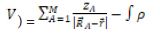
where ZA is the charge of nucleus A with position RA and p(r) is the electron density of the molecular system at the point, M is the number of nuclei in the system.The projections of molecular electrostatics potential (MESP) of keratin using Xcrysden and Jmol are given in Fig. 2(a,b,c,d). Molicular surface around keratin using Xcrysded and Jmol represented in Figure 2(a & b). Different colour on the surface of molecule corresponds to different values of electrostatic potential. Reds colour represents regions of most negative electrostatic potential, (i.e. oxygen) blue colour represents regions of most electro positive ESP (i.e. hydrogen) yellow colour around sulphur represent moderate electrostatic potential and green colour represents regions of zero potential (around carbon) [22-24]. The negative electrostatic potential (red regions) is localized over the oxygen atom that corresponds to an attraction of the proton by the aggregate electron density. The most positive regions (blue regions) correspond to proton repulsion (shaded as blue) are localized over hydrogen atoms. Figure 2(c) is Van der Waal's surface represented by ball and stick model, here sulphur (yellow), hydrogen (white) and oxygen is red [25-28] it shows that total electron density aroun
Charge density analysis
The nature of chemical bonds analysed by charge density distribution using Xcrysden. It can display large densities, molecular orbitals, or any other 2D or 3D scalar field in form of isosurface or contours. It require XSF file obtain by SIESTA output. Figure 3 (a,b,c,d) shows change density distribution of keratin at different resolutions [29,30]. It is observed from the figure that C-C and C-H bond are covalent in nature while C-O bond are partially convent and partially ionic but S-S bond are ionic in nature which leads to localized wave functions shown by concentric spheres centred at atoms with a smaller degree of overlap. The charge density distributions in the systems are higher in the inner-most region (magenta), and lower in the outer most regions (red). Higher charge density is observed around oxygen and sulphur because of higher value of ionization energy or electronegativity than other atoms present in compound. The concentric circles around oxygen and sulphur atoms represents ionic bond while distortion in circles between carbon and oxygen is an indication of a partial covalent and partial Ionics. The overlapping lobe between carbon nitrogen represent the bond is covalent (Figure 3).
Density of States and partial density of states Analysis
Number of different states at a particular energy level that electrons are allowed to occupy or electron states per unit volume per unit energy is classed Density of states (DOS). The projected or partial density of states (PDOS) is the relative contribution of a particular atom or orbital to the total DOS [31,32]. It provide quick qualitative picture of the electronic structure of a material. The density states (DOS) of keratin and projected density of states (PDOS) of different atoms contributed in DOS obtain by Siesta output are analysed by Xmgrace programme are presented in Figure 4 and in Figure 5 respectively.
From figure 4 is DOS of keratin, from this graph we can calculate both the frontier molecular orbitals i.e. highest occupied molecular orbital (HOMO) and lowest unoccupied molecular orbital (LUMO) taking part in chemical reaction. The HOMO is the outermost (highest energy) orbital containing electrons that could act as an electron donor while LUMO is the innermost (lowest energy) orbital that has room to accept electrons and can act as the electron acceptor. According to the frontier molecular orbital theory, the formation of a transition state is due to an interaction between the frontier orbitals (HOMO and LUMO) of reactants [33-35]. The energy value corresponding to LUMO and HOMO calculated by DOS analysis are found to be -1.24 eV and 1.56 eV respectively, the forbidden energy gap between valence and conduction band is about 3.8 eV which shows insulating behaviour of keratin. Below LUMO at energy level -3.2 eV and -4.20 eV some more peak are obtain which represent valence band completely filled by electron of different atoms similarly above HOMO the peaks represent electrons of atoms present in conduction band.
Contribution of different atoms in conduction band valence band analysed by Partial density of states (PDOS) which is shown in Figure 5. This figure represent that wave function of 2p orbitals of C, O, N and S and 2s orbital of N and O corresponding to energy value 1.24 eV overlap and form lowest unoccupied molecular orbital (LUMO). Highest occupied molecular orbital (HOMO) form by overlapping of 2p orbital of N and O and 2s orbital of oxygen at energy level 1.56 eV. It is also seen that in conduction band electrons of H(1s orbital) and O(2p orbital) play main role but in valence band electrons of C(2p orbital) and N(2p orbital) electrons are primary contributors (Figures 4 & 5).
Acknowledgments
Authors are grateful to Dr M L Verma (HOD, SSGI Bhilai), Dr B Keshev Rao (Professor SSGI, Bhilai) and to the manage- ment of Shri Shankaracharya Technical Campus-SSGI for providing lab facility to their campus and special guidance to lean SIESTA. I also thanks to Mr. Anand Tripathi (Chairman KEC, Bhilai), Mr. Abhishek Tripathi (Director KPSI, Raipur), Ms Vinita Mairal (Principal KPSI Raipur) and Ms Sumangla Sundri (Vice Principal KPSI Raipur) for providing overall support to complete this research work.
Conclusion
Keratin is capable to recover and/or improve the mechanical properties of hair. It is present in your skin, hair, and nails where they help to make these tissues strong and flexible. From DFT analysis of keratin it is concluded that band gap of keratin is 3.8 eV thus it has insulating nature. Molecular surface of keratin shows that there is no active site thus due to insulating nature and lacking of active site keratin protect our hair, skin and nails to react from outer atmospheric contamination. Ionic nature of bond and longest bond length between S-S atoms shows that there is weak attraction between them. Thus this bond is mainly cooperate in smoothness and curling nature of hair. Finlay it can be seen that S-S bond play main role in keratin for their strength. Thus for the strength of keratinocyte protein it should be advice to use sulphur rich oil or food. It can be obtain from animal and plant based protein as well as other types of compounds such as sulfinates, allicin, and sulphides. Sulphur is also present in thiamine (vitamin B-1) and biotin (vitamin H).
- Acda MN (2010) Waste chicken feather as reinforcement in cement-bonded composites. Philippine J Sci 139: 161-6.
- Agrahari S, Wadhwa N (2010) Degradation of chicken feather a poultry waste product by keratinolytic bacteria isolated from dumping site at Ghazipur poultry processing plant. Int J Poult Sci 9: 482-9.
- Alsarra IA (2009) Chitosan topical gel formulation in the management of burn wounds. Int J Biol Macromol 45: 16–21.
- Aluigi A, Sotgiu G, Ferroni C, Duchi S, Lucarelli E, et al. (2016) Chlorin e6 keratin nanoparticles for photodynamic anticancer therapy. RSC Adv 6: 33910-8.
- José M Soler, Emilio Artacho, Julian D Gale, Alberto García, Javir Junquera (2002) J Physics: Condensed Matter 14: 11.
- Franco Bassani, Gerald L. Liedl, Peter Wyder (2005) Encyclopedia of Condensed Matter Physics ReferenceWork ISBN 9780-12-369401-0.
- Vrugt Michael, Lowen Hartmut Wittkowski, Raphael (2020) Classicdynamical density functional theory: from fun- damentals to applications”. Advances in Physics 69: 121–247.
- Evans Robert Oettel, Martin Roth, Roland Kahl Gerhard (2016) New developments in classical density functional theory”. J Physics: Condensed Matter. 28: 240401.
- Martin RM (2004) Electronic Structure: Basic Theory and Practical Methods, Cambridge University Press.
- Artacho E, Cela JM, Gale JD, Garcia A, Junquera J, et al. (2011) J. Chem. Phys 152: 204108
- José MS, Emilio A, Julian DG, Alberto G, Javier J, et al. (2002) J Phys Condens Matter 14: 2745.
- Ordejón P, Artacho E, Soler JM (1996) Phys Rev B 53: 10441.
- Kang HT, Hwang ES (2006) 2-Deoxyglucose: An anticancer andantiviral therapeutic, but not any more a low glu- cose mimetic. Life Sci 78: 1392-9.
- Bharatam PV, Patel DS, Iqbal P (2005) Pharmacophoric featuresof biguanidienes: an electronic structure study. J Med Chem 48: 7615–22.
- Bharatam PV, Sundriyal S (2006) Molecular electrostatic poten-tials in the design of dendrimers for the delivery of glitazones. JNanosci Nanotechnol 6: 3277–82.
- Roy DK, Balanarayan P, Gadre SR (2009) Signatures ofmolecular recognition from the topography of electrostaticpotential. J Chem Sci 121: 815–21.
- Rastelli G, Pacchioni S, Sirawaraporn W, Sirawaraporn R, ParentiMD, Ferrari AM (2003) Docking and database screening revealnew classes ofP. falciparumdihydrofolate reductase inhibitors. JMed Chem 46: 2834-45.
- Hohenberg P & Kohn W (1964) Inhomogeneous Electron Gas, Phys. Rev. B, 136B: 864.
- Joshi BD, Srivastava A, Honorato SB, Tandon P, Pessoa ODL, et al. (2013) Study of molecular structure, vibrational, electronic and NMR spectra of oncocalyxone A using DFT and quantum chemical calculations, Spectrochim. Acta A 113: 367.
- Politzer P & Truhlar DG (1981) Chemical Applications of Atomic and Molecular Electrostatic Potentials, Plenum, New York, 1981.
- Pingale SS (2011) Molecular Electrostatic Potentials Concepts and Applications. Phys Chem 13: 15158.
- Politzer P, Grice ME, Murray JS, Seminario JM (1993) Anomalous Stabilizing and Destabilizing Effects in Some Cyclic p-Electron Systems, Can J Chem 71: 1123.
- Suresh CH, Alexander P, Vijayalakshmi KP, Sajith PK, Gadre SR (2008) Use of molecular electrostatic potential for quantitative assessment of inductive effect Phys Chem 10: 6492.
- Balnarayan P, Gadre SR (2003) Topography of molecular scalar filds. I. Algorithm and Poincare-Hopf relation, J Chem Phys 119: 5037.
- JM Soler, E Artacho, JD Gale, A García, J Junquera, et al. (2002) “The SIESTA method for ab initio ordern materials simulation. J Phys Condens Matter 14: 2745–79.
- E Artacho, E Anglada, O Diéguez, JD Gale, A García, et al. (2008) The SIESTA method; developments and applicability. J Phys Condens Matter 20: 064208.
- C Kittel (1986) Introduction to Solid State Physics (John Wiley & Sons, New York, 1986).
- L Lin, A García, G Huhs, C Yang (2014) SIESTA-PEXSI: Massively parallel method for efficient and accurate ab initio materials simulation without matrix diagonalization. J Phys Condens. Matter 26: 305503.
- Omprakash Verma, MR Meshram, AK Mishra, Mohan L, Verma B, et al. (2020) Structural, electronic and optical properties of In2O3: a density functional study, J Optical and Quantum electronics 5.
- Upma, Mohan L Verma (2019) Structures and Properties of Biopolymer Chitosan A First Principle Study, J. I manager.
- F Pauly Beilstein (2015) J Nanotechnol 6: 1247-59.
- Kaliginedi V, Moreno-García P, Valkenier H, Hong W, García-Suárez VM, et al. (2012) Wandlowski, T. J. Am. Chem. Soc 134: 5262-75.
- Perrin ML, Frisenda R, Koole M, Seldenthuis JS, Gil JAC et al. (2014) J. Nat. Nanotechnol 9: 830-4.
- Strange M, Thygesen KS, Beilstein (2011) J Nanotechnol 2: 746-54.
- Ning J, Li R, Shen X, Qian Z, Hou S, et al. (2007) Nanotechnology 18: 345203.
FIGURE 1
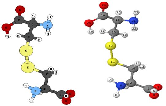
Figure 1: Optimized Structural of Keratin (a) With atomic symbol (b) With numbering of atom
FIGURE 2
.JPG)
.JPG)
Figure 2: (a) Molecular surface (b) Electrostatic potential surface (c) electron density and (d) molecular electrostatic
FIGURE 3
.JPG)
.JPG)
Figure 3: Charge density analysis of keratin at different magnification
FIGURE 4
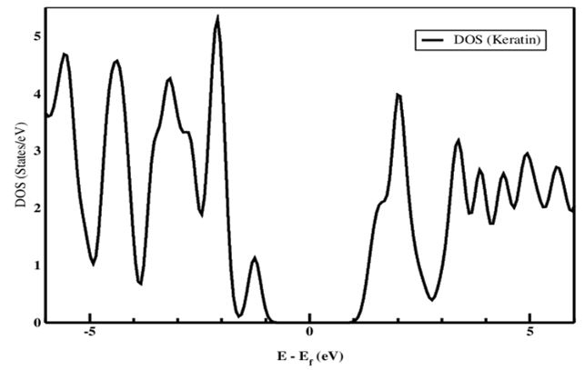
Figure 4: Density of State (DOS) of Keratin
FIGURE 5

Figure 5: Partial Density of State (PDOS) of Keratin
Tables at a glance
Figures at a glance