Optimized Hyper Heuristic Model for Efficient Diabetic Retinopathy Detection Using Deep Learning
Received Date: April 14, 2025 Accepted Date: May 14, 2025 Published Date: May 17, 2025
doi:10.17303/jaist.2025.2.102
Citation: M. Asif Shaikh, Syed Asif Ali (2025) Optimized Hyper Heuristic Model for Efficient Diabetic Retinopathy Detection Using Deep Learning. J Artif Intel Sost Comp Tech 2: 1-9
Abstract
This research aims to design an enhanced Diabetic Retinopathy Detection System using optimized Hyper Heuristic Model. With the growing significance of Diabetic Retinopathy Detection, there is an urgent need for an efficient model with superior accuracy.
Diabetic retinopathy is a diabetes associated difficulty that affects the eyes and can lead to vision loss or blindness if not treated immediately. Early detection is vital in preventing visual loss, reducing healthcare costs, microvascular complications like diabetic nephropathy or neuropathy. This study proposes optimized Hyper Heuristic Model based on Image Processing and Deep Learning algorithms, leveraging Genetic Algorithm, Convolutional Neural Networks, Histogram Equalization, Segmentation and Morphological Operations. A hyper heuristic model is a high-level approach in optimization and artificial intelligence that focuses on selecting, generating, or combining lower level heuristics to solve complex problems effectively. This research considers the strength of hyper heuristic methodologies to enhance diagnostic accuracy, reduce computational overhead, and support healthcare professionals in combating vision loss due to diabetic retinopathy. The research will focus on retinal images with the goal of describing accurate Diabetic Retinopathy. By conducting these experiments and analyses, the study intends to provide accurate, efficient and reliable Diabetic Retinopathy results of different images from different datasets to provide a comprehensive solution. The findings from this research are expected to helpful in detection of early Diabetic Retinopathy and preventing possible vision loss in diabetic patients. This work will contribute to Eye Specialist, Dialectologists and Diabetic Patients providing new insights into Diabetic Retinopathy Detection.
Keywords: Diabetic; Retinopathy; Retinopathy Detection System; Hyper-Heuristic Model (HHM) Convolutional Neural Networks (CNN); Machine Learning
Introduction
A. Diabetic means anything related to diabetes, which is a chronic medical condition that is categorized by the sugar levels also called glucose in the blood [1]. There are different types of diabetes, including:
Type 1 Diabetes: It is an automatic immunity situation when the body's immune system attacks the pancreas cells that produce insulin. This fallout in little to no insulin production, which forces individuals to take insulin injections.
Type 2 Diabetes: A situation where the body becomes resilient to insulin or doesn't produce enough insulin. It is often related with lifestyle factors, such as obesity and physical dormancy, and is more common in adults.
Gestational Diabetes: It is a type of diabetes that arises during pregnancy. It usually resolves after childbirth but rises the risk of developing type 2 diabetes later in life.
B. Retina is a thin layer of tissue that is located at the back of an eye. It is a crucial part of the eye's anatomy because it produces electrical signals from light that enters the eye, and then an optic nerve sends this to brain. The brain reads these signals to generate the images we see [2]. The retina is vital for vision, and damage to it can lead to serious visual impairments. Conditions like diabetic retinopathy, macular degeneration, and retinal detachment can all affect the retina and potentially lead to blindness if not treated. Regular eye examinations are important for detecting and managing retinal conditions.
C. Microaneurysms are tiny, balloon like swellings that occur in the walls of the capillaries which are small blood vessels in the retina, a light sensitive tissue at the back of the eye. These are among the earliest visible signs of diabetic retinopathy, an impediment of diabetes that affects the eyes [3].
D. Hyper Heuristic Models are advanced strategies used in optimization that operate at a higher level than traditional heuristics. Instead of solving a specific problem directly, hyper-heuristics focus on selecting, generating, or adapting low level heuristics to enhance the performance of solving complex problems. They aim to automate and enhance the process of heuristic application and design [4].
E. Image Processing are techniques to improve the quality of images, extracting useful information, or prepare them for further analysis. This includes both digital images and video. The goal of image processing can range from enhancing image quality to extracting meaningful data from images [5].
Research Background
The summary of research papers covers different approaches and methodologies for the detection of Diabetic Retinopathy (DR) and Microaneurysms (MAs) using various techniques that includes Image Processing, Deep Learning and Evolutionary Algorithms.
An advance deep learning approach proposes ResNet50 for diabetic retinopathy detection and classification with an accuracy of 87%, precision 84% and F1 val- ue 86 [6].
A hybrid neural network approach for classifying diabetic retinopathy and its sub- types is proposed using EfficientNet and Swin upon APTOS-19 with an accuracy of 97% [7].
Two methods each with two systems to detect the developmental stages of DR using fundus images was proposed [8] where in first method, GoogLeNet and Res- Net-18 with SVM are used while in second method, Feed Forward Neural Networks based on the hybrid features ex- traction using GoogLeNet Fuzzy color Histogram, Gray Lev- el Co=occurrence Matrix, Local Binary Pattern and Res- Net-18 are used. This method has obtained 99.7% accuracy, 99.6% precision, 99.6% sensitivity, 100% specificity, and 99.86% AUC.
A deep learning model was used in a real time scenario of Sindh Institute of Ophthalmology & Visual Sciences (SIOVS). Test images quality was assessed by classifying them in DR Positive and DR Negative. Specialists studied results of 398 patients for five weeks, in those patients there were 232 males and 166 fe- males and accuracy of 97.30% was achieved [9].
Deep learning-based metaheuristic model for diagnosing of DR proposed a method uses preprocessed retinal images, extracts features by using InceptionV3 which is a deep learning model, classification and feature selection with XGBoost and Simulated Annealing. Accuracy of 92.55% was achieved on Messidor-2 dataset. This study com- pares suggested hybrid model with other models using same datasets [10].
A hybrid model for the identification and classification of Diabetic Retinopathy stages was put forward [11]. The model uses Image Preprocessing, Local Binary Patterns, collaborative and Deep Learning features that deliver vector optimized based collective feature on the Binary Dragonfly Algorithm and the Sine Cosine Algorithm.
A model focuses on the detection and classification of Diabetic Retinopathy using a technique based on feed forward neural networks, which focus on green channel extraction, histogram equalization, and extracting candidate region [12].
[7] proposes a Convolution Neural Networks for diabetic retinopathy identification using a segment-based learning approach on fundus images. It comprises adaptation of a pre-trained Convolution Neural Networks and integration of all segment levels of the diabetic retinopathy map for learning image level classifiers and features
A model uses descriptive statistics and machine learning methods to examine the identification of microaneurysms in diabetic retinopathy. Familiar machine learning approaches with a deep learning-based method for MA detection are also compared [14].
A Convolution Neural Networks for diabetic retinopathy identification using a segment-based learning approach on fundus images was proposed [13].
A segment based learning approach suggests that significant improvements in recognizing of diabetic retinopathy images and their lesions is possible, involving the adaptation of pre-trained CNNs for the segment level assessment of diabetic retinopathy and the integration of all segment levels for image classification [15].
An automatic method for identifying microaneurysms in fundus photographs using a convolutio- nal neural network with batch normalization and Dice loss was proposed, achieving better results than state of the art methods by the free response receiver operating characteristic (FROC) metric [16].
A hybrid feature implanting approach utilizes pre-trained CNN models for early detection of MAs, with performance evaluations on openly available datasets "E- Ophtha" and "DIARETDB1” [17].
Research Outcomes
Some model studied emphasis on DR detection and classification [12,13], some precisely address microaneurysm detection [14-17]. Some gives high accuracy in the detection of DR, achieving 98.89% and 96.97% accuracy, respectively, representing the effectiveness of their proposed methods [14-17]. However, [16,17] concentrate on microaneurysm detection, reaching good results than state of the art methods.
Two model were compared for traditional machine learning methods with deep learning-based methods for MA detection [14,15], demonstrating the superiority of old style machine learning methods over deep learning-based MA detection.
Two model propose hybrid Meta heuristic techniques based upon deep learning algorithms, where earlier gives an accuracy of 99.7% on the Kaggle while latest gives 92.55 on Messidor-2 dataset [10,18].
A model proposes approaches for DR detection and classification where earlier gives accuracy of 87% and later gives 97% [6].
Differences in Methodologies and Focus
Overall, the methodologies in the papers differ in terms of the specific detection target (DR vs. MAs), approach (machine learning vs. deep learning), and the evaluation of results. While some papers focus on improving the accuracy of DR detection, others emphasize the superiority of traditional machine learning methods over deep learning in MA detection.
Limitations
Complexity of Retinal Images: Retinal images are complex, with various features such as blood vessels, optic discs, and pathological lesions (e.g., microaneurysms, hemorrhages) that need to be distinguished for accurate diagnosis.
Early Detection: Early-stage DR is characterized by subtle features like microaneurysms, which are small and difficult to detect, leading to potential under diagnosis.
Large Scale Screening: The need for large scale screening in diabetic people requires automated systems that can process thousands of images accurately and efficiently.
Variability in Image Quality: Retinal images can vary meaningfully in quality due to features like lighting situations, patient movement, and equipment used, which can affect the accuracy of detection algorithms.
Proposed Methodologies
Algorithm of Optimized HHM for DR Detection
1) Preprocessing
a) Input Normalization: Normalize the intensity values of images to a standard scale
b) Noise Reduction: Apply Gaussian filtering to reduce image noise while preserving edges.
c) Contrast Enhancement: Use histogram equalization to enhance visibility of retinal features.
d) Region of Interest Extraction: Focus on retinal regions by detecting and cropping the optic disc and macula using morphological operations.
2) Feature Extraction
a) Texture Features: Extract texture features using GLCM (Gray-Level Co-Occurrence Matrix).
b) Color Features: Extract color histograms to capture hemorrhages and exudates.
c) Deep Features: Pass the preprocessed images through a pre-trained CNN and extract feature vectors from the last convolutional layer.
3) Multi Model Fusion
a) Combine Features: Concatenate texture, color, and deep features into a single feature vector
b) Dimensionality Reduction: Use Principal Component Analysis to reduce feature dimensionality while retaining significant information.
4) Hyper Heuristic Model Optimization
a)Objective Function: Define the detection model's objective function which minimizes the loss.
b)Search Space Definition: Define heuristics to initialize search space (feature selection, hyper parameters etc.)
c)Meta Heuristic Optimization: Use algorithms like Genetic Algorithms or Particle Swarm Optimization to explore and exploit the search space. Evaluate fitness for each heuristic combination using cross validation.
5) Classification
a) Train a deep learning classifier: Fine-tuned CNN on the optimized features.
b) Predict Classes: Output the predicted class probabilities and apply thresholding for classification into DR severity levels.
6) Post-Processing
Refine predictions using domain knowledge using spatial distribution of lesions.
7) Validation and Testing
a)Model Evaluation: Evaluate the model using metrics like accuracy, precision, recall, F1-score, and AUC-ROC and Return final predictions.
B. Datasets
Following datasets will be used to train the model. These are the most commonly used and highly regarded retinal image datasets for research in diabetic retinopathy detection and other retinal diseases:
Kaggle Diabetic Retinopathy Detection Dataset:
OCTID
Results
- Description: One of the largest publicly available datasets for diabetic retinopathy detection. It covers high resolution retina images taken under various imaging conditions. The images are labeled with a grade indicating the severity of diabetic retinopathy.
- Size: 88,702 images
- Use Cases: Diabetic retinopathy detection, classification tasks
- Description: A dataset emphasized on Optical Coherence Tomography (OCT) images. It is used in research for segmenting retinal layers and identifying conditions like macular degeneration.
- Size: 269,000 images (from 1,600 patients)
- Use Cases: OCT image analysis.
The proposed Optimized Hyper Heuristic Model (HHM) for diabetic retinopathy (DR) detection integrates image processing techniques, deep learning frameworks, and metaheuristic optimization to achieve exceptional accuracy and efficiency. Although experimental results are pending, the model is projected to attain an accuracy of 98–99%, with precision and recall metrics surpassing 97%, through the strategic integration of handcrafted and deep features optimized via methods such as Genetic Algorithms and Principal Component Analysis (PCA). Its robust preprocessing pipeline significantly enhances image quality, facilitating the effective early detection of DR markers, notably microaneurysms. Additionally, the hyper-heuristic component automates parameter tuning, promoting adaptability across various datasets, including Kaggle DR and OCTID. Once validated, the HHM is expected to outperform traditional methods, delivering a scalable and highly accurate solution suitable for practical DR diagnosis applications.
Conclusion
The proposed Optimized Hyper Heuristic Model (HHM) demonstrates a novel and robust approach to accurate and efficient diabetic retinopathy (DR) detection by integrating traditional image processing techniques, deep learning algorithms, and a hyper-heuristic optimization framework. This hybrid methodology leverages the strengths of handcrafted feature extraction (texture and color analysis) and deep features from CNN architectures, achieving a comprehensive representation of retinal abnormalities.
As this paper presents a theoretical model, no experimental results are included at this stage; however, comprehensive results will be provided in forthcoming publications
The hyper-heuristic layer ensures dynamic adaptability, optimizing the detection pipeline by automating the selection of preprocessing steps, feature extraction methods, and model parameters. This adaptability will not only improve detection accuracy across diverse images but will also reduce manual intervention, making the model scalable for real-world applications.
By combining accuracy, efficiency, and interpretability, the HHM will provide a significant advancement in diabetic retinopathy detection. It supports early diagnosis and timely treatment, potentially reducing the global burden of diabetes-related vision loss. Future work could focus on extending the model to multi-disease detection and deploying it in real-time clinical environments to further enhance its impact.
- Singh A, Shadangi S, Gupta PK, Rana S (2025) Type 2 Diabetes mellitus: A Comprehensive review of pathophysiology, comorbidities, and emerging therapies. Comprehensive Physiology, 15.
- Abramoff MD, Garvin MK, Sonka M (2010) Retinal Imaging and Image Analysis. IEEE Reviews in Biomedical Engineering, 3: 169-208.
- Mayya V, Kamath S, Kulkarni U (2021) Automated microaneurysms detection for early diagnosis of diabetic retinopathy: A Comprehensive review. Computer Methods and Programs in Biomedicine Update, 1: 100013.
- Özcan E, Bilgin B, Korkmaz EE (2008) A comprehensive analysis of hyper-heuristics. Intelligent Data Analysis, 12: 3-23.
- Visual research. (n.d.). Google Books. https://books.google.com.pk/books?hl=en&lr=&id=yaRLAQAAQBAJ&oi=fnd&pg=PR5&dq=The+goal+of+image+processing+can+range+from+enhancing+image+quality+to+extracting+meaningful+data+from+images+(Marion,+2013).&ots=1c9550TelQ&sig=vzpm24q1nVCpdyDZ_ekCN0LoFLM&redir_esc=y#v=onepage&q&f=false
- Khalil A, Khan FU, Faisal M, Hasanat M, Jan A, Ali Y. Advance Deep Learning Approach for Diabetic Retinopathy Detection and Severity Classification Utilizing ResNet50.
- Xu X, Zhang M, Huang S, Li X, Kui X, Liu J (2024) The application of artificial intelligence in diabetic retinopathy: progress and prospects. Frontiers in Cell and Developmental Biology, 12.
- Alshahrani M, Al-Jabbar M, Senan EM, Ahmed IA, Saif JaM (2023) Hybrid Methods for Fundus Image Analysis for diagnosis of Diabetic Retinopathy Development stages based on fusion features. Diagnostics, 13: 2783.
- Bajwa A, Nosheen N, Talpur KI, Akram S (2023) A Prospective Study on Diabetic Retinopathy Detection Based on Modify Convolutional Neural Network Using Fundus Images at Sindh Institute of Ophthalmology & Visual Sciences. Diagnostics, 13: 393.
- GÜRCAN ÖF, ATICI U, BEYCA ÖF (2022) A Hybrid Deep Learning-Metaheuristic Model for Diagnosis of Diabetic Retinopathy. GAZI UNIVERSITY JOURNAL of SCIENCE.
- Ishtiaq U, Abdullah ERMF, Ishtiaque Z (2023) A hybrid technique for diabetic retinopathy detection based on Ensemble-Optimized CNN and texture features. Diagnostics, 13: 1816.
- R Deepa, Narayanan NK (2020) Detection of Microaneurysm in Retina Image using Machine Learning Approach.
- Math L, Fatima R (2020) Adaptive machine learning classification for diabetic retinopathy. Multimedia Tools and Applications.
- Cao W, Czarnek N, Shan J, Li L (2018) Microaneurysm detection using principal component analysis and machine learning methods. IEEE transactions on nanobioscience, 17: 191-8.
- Math L, Fatima R (2019) Identification of diabetic retinopathy from fundus images using CNNs. International Journal of Innovative Technology and Exploring Engineering, 9: 3439-43.
- Chudzik P, Majumdar S, Calivá F, Al-Diri B, Hunter A (2018) Microaneurysm detection using fully convolutional neural networks. Computer methods and programs in biomedicine, 158: 185-92.
- Mateen M, Malik TS, Hayat S, Hameed M, Sun S, Wen J (2022) Deep Learning Approach for Automatic Microaneurysms Detection. Sensors, 22: 542.
- Uzair Ishtiaq, Rahayu E, Zubair Ishtiaque (2023) A Hybrid Technique for Diabetic Retinopathy Detection Based on Ensemble-Optimized CNN and Texture Features. Diagnostics, 13: 1816.
FIGURE 1
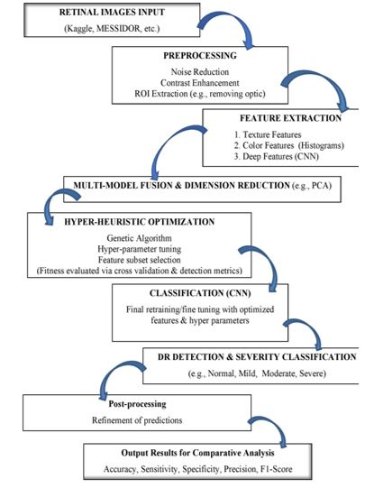
Figure 1: Conceptual Diagram for Algorithm of Optimized HHM for DR Detection
FIGURE 2
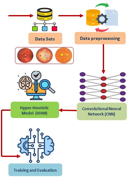
Figure 2: Data Processing Diagram
Tables at a glance
Figures at a glance