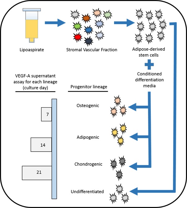
Figure 1 Study design: Beginning with fresh lipoaspirate, SVF was enzymatically isolated and the ASCs were expanded after plastic adherence. The ASCs were then split into three differentiation media for osteogenic, adipogenic, and chondrogenic progenitor development. The supernatant of each lineage was then sampled on culture days 7, 14, and 21 and the sVEGF-A165 quantities were determined by immunoassay. Similarly, first passaged undifferentiated ASCs were also expanded and comparatively tested.
Figure 2 Undifferentiated adipose-derived stem cells.
Representative bright-field 10x micrograph illustrating plastic plate adherence and outward extension of non-passaged adipose-derived stem cells (undifferentiated growth day 10).
Figure 3Figure 3. Adult progenitor cells, differentiation day 14. (A) Representative bright-field 15x micrograph illustrating calcium staining by Alizarin Red of adipose-derived stem cells differentiated to osteoblasts. (B) Representative bright-field 30x micrograph illustrating lipid droplet staining of adipose-derived stem cells differentiated to adipocytes. (C) Representative indirect micrograph illustrating a sulfated proteoglycan sphere stained with alcian blue & nuclear fast red indicating adipose-derived stem cells differentiated to chondroblasts. Sphere diameter 2mm.
Figure 4 Osteogenic progenitor cell colony
Alizarin Red staining alone for calcium confirms a functional osteogenic progenitor cell colony (40x magnification).
Figure 5 Osteogenic progenitor sphere stained for vegf-a165.
(A) Presumed osteogenic progenitor sphere (ASCs treated with osteogenic differentiation media), unstained, 20x mag. (B) Same presumed osteogenic progenitor sphere positively stained with DAB (dark brown) alone, indicating the production of VEGF-A165, 20x mag.
Figure 6 Osteogenic progenitor cell sphere, stained with alizarin red and dab.
This osteogenic progenitor sphere has been stained with Alizarin Red (for calcium) and DAB (for VEGF-A165), 20x mag. The staining is comparatively lighter than with DAB alone and darker than Alizarin Red alone, diagnostic for an osteogenic progenitor sphere producing VEGF-A165.
Figure 7 Secreted Vegf-A165 Levels (means of the three cell lines respectively).
At days 7, 14, and 21 of lineage-specific culture, samples of each APC supernatant (osteogenic, adipogenic, and chondrogenic) were obtained and VEGF-A165 levels determined by immunoassay. Similarly, the VEGF-A165 levels of undifferentiated first passaged ASCs were also measured. The osteogenic media displayed the highest levels of VEGF-A165. The chondrogenic lineage secreted the least amount of VEGF-A165.
Figure 8 Secreted Vegf-A165 Levels (by cell line)
Each of the three cell lines produced respectively different quantities of VEGF-A165, through respective levels trended similarly: panel A (osteogenic APCs), panel B (adipogenic APCs), panel C (ASCs), panel D (chondrogenic APCs).
Figure 9 Secreted vegf-a165 maximum levels per 106 adipose-derived progenitor cells.
These values are reflective the maximal quantity of VEGF-A165 as sampled on differentiation day 14(osteogenic, adipogenic, and chondrogenic APCs) and culture day 21 for undifferentiated ASCs.
Figure 10 Comparative quantities of apcs to achieve vegf-a165 ed50 for human Umbilical vein endothelial cell (huvec) stimulation. Correlation is specific to Current Good Manufacturing Practice recombinant VEGF-A165 (Cat# 293GMP, R&D Systems, Minneapolis, MN) and secreted VEGF-A165 levels are reflective of maximum sampling lots (day 14 for APCs and day 21 for undifferentiated ACSs).
Figure 11 Central Illustration: In this photomicrograph, this osteogenic progenitor cell sphere has been stained for both calcium (confirming osteogenic function) as well as VEGF, confirming that the osteogenic progenitor cells are indeed producing both calcium and VEGF.