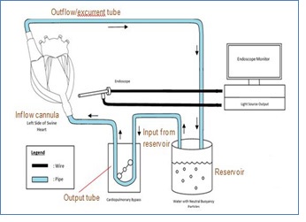
Figure 1:Schematic diagram reflecting the overall Set-up
Figure 1:Schematic diagram reflecting the overall Set-up

Figure 2:Operative set-up of radioscopic evaluation of explanted swine heart

Figure 3: Parachuting of the Valve into the Left Cardiac Chamber

Figure 4:(A) Cardioscopy Snapshot of Particle Movement at Respective Valves (B) Traced Outline of Particle Trajectory at Respective Valves. A1, B1: Native valve, A2, B2: Medtronic Open Pivot Mechanical Valve and A3, B3: Medtronic Mosaic Bioprosthetic Valve

Figure 5: Schematic of Traced Flow Pattern in Respective Valves. A: Native valve, B: Medtronic Open Pivot Mechanical Valve and C: Medtronic Mosaic Bioprosthetic Valve

Figure 6: (A) Initial Echocardiography to Visualise Flow Pattern and (B) Initial Trial of Cardioscopy to evaluate mitral valve morphology
Figures at a glance