Development and Characterisation of Ayurvedic Polyherbal Formulation for Diabetic Wound Healing: A Comprehensive Study on Decoction, Phytochemical Analysis, and Topical Application
Received Date: May 05, 2023 Accepted Date: June 05, 2023 Published Date: June 08, 2023
doi: 10.17303/jber.2023.7.101
Citation: Shivani Tank, Harishkumar Madhyastha, Shivani Patel, Anmol kumar (2023) Development and Characterisation of Ayurvedic Polyherbal Formulation for Diabetic Wound Healing: A Comprehensive Study on Decoction, Phytochemical Analysis, and Topical Application. J Biomed Eng Res 7: 1-17
Abstract
Diabetic wounds represent a significant challenge in modern healthcare, with delayed healing and increased susceptibility to infections. This study aims to synthesise and characterise an Ayurvedic polyherbal formulation from Securinega leucopyrus (Katupila), Azadiracta indica (Limbdo), Acacia catechu (Khadir) and Vitex negundo (Nirgundi) for diabetic wound healing. The research encompasses preparation by decoction, followed by qualitative and quantitative phytochemical analysis, and biological assays including antioxidant, and antibiofilm activity against pathogens. Additionally, the study examines the development of a topical ointment for improved application on diabetes mellitus patients' wounds. The isolation and characterization of bioactive compounds are conducted using GCMS techniques.
Keywords: Diabetic wound healing; Amputation; Phytochemicals; Bio-actives
Introduction
Diabetic Wound Healing ChallengesDiabetes is the aggregation of complications at metabolic state involving a myriad of comorbidities including the serious conditions of poor wound healing, chronic ulceration, neuropathy, ischemia, and immune dysfunction and resultant amputation of limb. The epidermal wound healing is a definite and orderly phase while the diabetic condition makes it distorted at all stages. While the etiology of chronic, non-healing category, diabetic wounds is multi-- faceted leading to the progression to a non-healing phenotype that has been quite linked to poor vascular networks [32]. Diabetes mellitus (DM) is a chronic metabolic disorder characterized by hyperglycaemia, which can lead to various complications, including impaired wound healing. Diabetic wounds often exhibit delayed healing, increased susceptibility to infection, and a higher risk of amputation. Conventional wound care management strategies have proven insufficient in addressing these challenges, necessitating alternative therapeutic approaches. DM is associated with various long-term complications affecting multiple organ systems, including the cardiovascular, renal, nervous, and integumentary systems. These complications can lead to significant morbidity and mortality and impose a substantial socioeconomic burden. Among these complications, diabetic foot ulcers (DFUs) are a significant challenge that affects approximately 15% of individuals with DM during their lifetime.
Ayurveda and Polyherbal FormulationsAyurveda, an ancient Indian system of medicine, has been used for thousands of years to treat various ailments. Ayurvedic polyherbal formulations are mixtures of multiple medicinal plants, which are believed to have synergistic effects that can enhance therapeutic benefits. These formulations are known for their multi- targeted actions, fewer side effects, and holistic approach to health. Polyherbal formulations have been reported to exhibit anti-inflammatory, antioxidant, and antimicrobial properties, which could contribute to wound healing in diabetic patients.
Materials and Methods
Plant materials and sample preparationPlant materials of Certain xerophytic plants undertaken for research are Securinega leucopyrus [69], Vitex negundo [74], Acacia catechu [83] and Azadiracta indica [85], collected from Saurashtra region, and their botanical identities were confirmed by a certified botanist Dr. Neha Patel, Atmiya University. The plant samples were washed, air-- dried under shade, and then ground into a fine powder using a mechanical grinder. The powdered samples were stored in air-tight containers until further use
Preparation of the Ayurvedic polyherbal formulationThe Ayurvedic polyherbal formulation was prepared through decoction, a traditional method of extracting bioactive compounds from plant materials. A specific ratio of the powdered plant materials was mixed and boiled in distilled water until the volume reduced to one-fourth. The decoction was then filtered and concentrated using a rotary evaporator to obtain the final extract [55,69].
Phytochemical analysis Qualitative phytochemical analysisThe qualitative phytochemical analysis of the polyherbal formulation was conducted using standard methods to identify the presence of various phytoconstituents, such as alkaloids, flavonoids, tannins, saponins, and terpenoids [7,27,89].
Quantitative phytochemical analysisQuantitative phytochemical analysis was performed to determine the total phenolic content, total flavonoid content, and total tannin content of the polyherbal formulation using spectrophotometric methods and standard calibration curves [7,27,89].
Extraction of phytochemicalsPhytochemical extracts were prepared for obtaining the bioactive compounds from the powdered mixture. For extraction of different bioactive compounds different solvents were used. These solvent were heated at their specific boiling points. The process involved different solvents like two polar solvents methanol & water, two semi- polar solvents chloroform & hexane and two non- polar solvents ethanol & petroleum ether. These extracts would be stored for further characterisation studies aimed to isolate the bioactive compounds.
Antioxidant assayThe antioxidant activity of the polyherbal formulation was evaluated using multiple assays, including DPPH (2,2-diphenyl-1-picrylhydrazyl) radical scavenging assay. The DPPH (2,2-diphenyl 1-picrylhydrazyl) assay was carried out according to the standard method. An aliquot of carotenoids (100μl) and equal volume of acetone and methanol were added to 950μl of 100μM DPPH methanol solution. The mixture was shaken vigorously and then left to stand at RT for 30 min in the dark [37]. The absorbance was measured spectrophotometrically at 580nm against an acetone/methanol (1/1, v/v) blank.
GCMS analysisControl absorbance - sample absorbance× 100 Gas chromatography-mass spectrometry (GCMS) analysis was employed to further elucidate the volatile compounds present in the polyherbal formulation. The analysis was conducted using a gas chromatograph equipped with a mass selective detector. The resulting data were used to identify and characterize the volatile compounds based on their retention time, mass spectra, and comparison with reference libraries.
Molecular Biology assayThe mouse skin fibroblast cell line m5S (4×10⁴ cells per mL) were cultured using medium α MEM supplemented with fetal bovine serum (10%) and antibacterial cocktail (1%) at room temperature (37 °C), with a continuous supply of CO2 and with maintenance of 95% humidity.
Cell Proliferation Assay by MTT was used for study of cell proliferation. Untreated cells were taken as Control. Cells were seeded in 96 well plates at a density of 1×10⁴ cells were treated with different doses of formulation for normal cell, for hyperglycaemic cell 0.5µl/ml, 1µl/ml in oil, 150µl/ml. Treated cells were washed with phosphate buffered solution (PBS). 100µl MTT solution was added into each well, and cells were incubated at 370 C for 4 hr. The resulting intracellular purple formazon was quantified with a spectrophotometer at an absorbance of 570nm (Multiskan FC, Thermo Fischer Scientific, Inc., Pittsburgh, PA, USA)
Quantification of intracellular reactive oxygen species (ROS) assayIntracellular ROS was quantified by using OxiselectTM Intracellular ROS assay kit (DoJindo, Inc, Washington, DC, USA). Cells were seeded on coverslips in 6- well plates at a density of 3×10⁵ cells/well. Cells were washed with PBS solution, 25µM DCFH-DA was added to cells 1 hr prior to treatment and incubated at 37°C. After incubation formulation was added on the basis of Cell Proliferation Assay (dose dependent) 30µl for normal cell and for hyperglycaemic cell Formulation doses were 0.5µl, 1µl and then incubated at 37°C for 2 and 4 hr. After treatment coverslip was removed in separate well washed with PBS solution 1-2 times. The cells were fixed on coverslip with 4%PFA or 70% Slides were mounted and images were captured using a confocal and fluorescence microscope (BZ-9000, Keyence, Osaka, Japan).
Cell Proliferation by Flow CytometerCell Apoptosis was quantified by an Annexin V-- FITC apoptosis detection kit (Nacalai Tesque, Tokyo, Japan). Normal and hyperglycaemic cells were seeded in 6- well plate density at – cells/well in the semi confluent phase were treated with varied concentration of Formulation for 2 and 4 hr. Cells were washed with PBS solution. Trypsin-EDTA was to for detach the cell from surface. Normal and hyperglycaemic medium was added in normal and hyperglycaemic cell respectively. Annexin binding buffer was added in the cell suspension, and then incubated with Annexin conjugate at room temperature for 15 min in the dark. Stained cells were added to PI (Propidium Iodide) solution before analyzed by Flow cytometry machine (BD Biosciences 7000)
Phagocytosis AssayPhagocytosis was determined using FITC (fluores-cein-isothiocyanate) phagocytosis kit (Cayman chemical, Ann Arbor, MI, USA). Hyperglycaemic cells seeded in 12- well plates at a 1×10 4 densities of cells/well were treated with formulation and for 2hr and 4hr and LPS was added for 4hr. After LPS treatment, cells were grown in hyperglycaemic DMEM medium for 24hr. After 24 hr, the cells were washed with PBS, and incubated with rabbit IgG-FITC conjugates latex beads for 3 hr. Nuclei were counterstained with DAPI and fluorescence images were obtained using confocal laser scanning microscope TCS SP8 (Leica, Germany).
In vitro cell migration assayIn vitro wound healing assay determined by using µ-well culture inserts (Ibidi Suppliers, Lochhamer, Grafelfing, Germany). Skin fibroblast cells (M5s) were culture in µwell with normal medium and glucose medium culture insert to fully confluent stage. Cells were treated with different concentration of formulation and for 2 hr and 4 hr incubated at 37°C. After 2 hr and 4 hr insert were slowly removed without disturbing the edge and cells were fixed with 100% Methanol for 10 min in room temperature. Fixed cells were washed with PBS and stained with 0.5% crystal violet cell staining dye for 5 min. Excess stain was removed by washing continuously with distilled water. Air dry the µ-well in aseptic condition. Photographs taken in (IX71, OLYMPUS, Japan).
Live Cell Migration AssaySkin fibroblast cells (M5s) were culture in µ-well culture to fully confluent stage. Inserted slowly and removed without disturbing the edge. Cells were cultured in standard cell culture medium with normal and glucose medium varied concentration of Formulation and in α MEM conditioned with at cell culture chamber. Live cell migration during different treatment was captured by time lapse video device (Cyto smart-II, Lonza, Inc., Morristown, NJ, USA) in conjunction with Image J (NIH) software.
Results and Discussion
Phytochemical analysis resultsThe qualitative phytochemical analysis of the Ayurvedic polyherbal formulation revealed the presence of various bioactive constituents, such as alkaloids, flavonoids, tannins, saponins, and terpenoids. The quantitative analysis showed significant levels of total phenolic content, total flavonoid content, and total tannin content, indicating the potential therapeutic properties of the formulation.
Antioxidant assayThe polyherbal formulation exhibited potent antioxidant activity in the DPPH assay suggesting its potential role in mitigating oxidative stress in diabetic wounds thereby indicating its potential to prevent and disrupt biofilm formation by pathogens, which could further enhance its therapeutic efficacy in diabetic wound healing.
Isolation and characterisation of bioactive compoundsGCMS analyses allowed for the identification and characterization of multiple bioactive compounds present in the polyherbal formulation. Some of these compounds have been previously reported to exhibit antioxidant, antiinflammatory, antimicrobial, and wound healing properties, further supporting the observed biological activities of the formulation. The identification of these bioactive compounds provides valuable insights into the potential mechanisms underlying the therapeutic effects of the Ayurvedic polyherbal formulation in diabetic wound healing.
Cell line analysis Cell proliferation assay: Dose and Time dependent assayMTT assay using for effect of Formulationand Kwath (aqueous extract) on normal and hyperglycaemic cell proliferation. Cells were treated with varied concentration of Formulation for 16hr. Normal and hyperglycaemic cell proliferation was higher in 30µl Formulation (Fig.6) in comparison to 150µl control. Examined the time course effect of 30µl Formulation (Fig.8) on Normal cell proliferation result showed that 2 hr when exposure to 30µl formulation and in hyperglycaemic cell examined time course effect of 0.5µl, 1µl and 150µl of formulation result showed that 4 hr caused a significant (p< 0.05) inhibition in cell proliferation.
Cell Apoptosis and Necrosis analysis by Annexin V-- FITC Apoptosis detection (FACS)The data obtained by flow cytometry assay shows the treatment with formulation to the normal cell exhibited % of live cells 94.70% which is almost same as control cells 95.03%. This suggest that the treatment with formulation is not toxic to the normal cells. In contrast when the hyperglycaemic cells were treated with formulation, the results showed % of live cells 75.93%, respectively which is significantly less toxic compared to control 61.30%.
Phagocytosis AssayPhagocytosis assay shows the treatment with formulation (0.5µl, 1µl) on macrophages cultivated high glucose α-DMEM media. cell compare to control the intensity of fluorescence is high.
In vitro Cell Migration AssayIn vitro cell migration assay shows that formulation accelerated cell migration in normal cell and hyperglycaemic cell in 18 hr. Observation showed cell elongation of dendrites in fibroblast and migration of border cells are indicative of invasiveness. Thus, the formulation proves to be effective for the cell migration and filling of gap was evident both in normal as well as hyperglycaemic cells.
Cell migration Assay in Normal cell lines Cell migration Assay in Normal cell lines Cell migration Assay in Hyperglycemic cell linesDiscussion
Phytochemical composition and its role in wound healingThe phytochemical composition of the Ayurvedic polyherbal formulation is rich in various bioactive constituents, such as alkaloids, flavonoids, tannins, saponins, and terpenoids. These phytoconstituents have been reported to possess multiple therapeutic properties that can contribute to the wound healing process. For instance, flavonoids and tannins are known for their antioxidant and anti- inflammatory activities, which can help alleviate oxidative stress and inflammation in diabetic wounds. Alkaloids and terpenoids have demonstrated antimicrobial properties, which can assist in controlling wound infections. Collectively, the presence of these bioactive constituents in the formulation could play a crucial role in promoting diabetic wound healing.
Biological activity and potential mechanisms of actionThe Ayurvedic polyherbal formulation exhibited potent antioxidant, antimicrobial, and antibiofilm activities, which can be attributed to the synergistic effects of its phytochemical constituents. The antioxidant activity of the formulation may help reduce oxidative stress in the wound environment, thereby promoting the healing process.
4.4 Bioactive compounds and their contribution to the overall activity of the formulationGCMS analyses identified several bioactive compounds present in the polyherbal formulation that may contribute to its therapeutic effects. These compounds have been reported to possess antioxidant, anti-inflammatory, antimicrobial, and wound healing properties. The presence of multiple bioactive compounds in the formulation suggests that its therapeutic potential could be attributed to the combined and synergistic actions of these constituents. This multi-targeted approach may offer a more comprehensive and effective strategy for diabetic wound healing compared to single-target therapies.
Conclusion
This study presents a comprehensive investigation into the synthesis, characterization, and evaluation of an Ayurvedic polyherbal formulation for diabetic wound healing. The phytochemical analysis revealed a rich composition of bioactive constituents, which contributed to the potent antioxidant activity observed. The formulation's efficacy in diabetic wound healing was further demonstrated through the development of a topical ointment and its evaluation in diabetic animal models.
The identification of bioactive compounds via GCMS analyses provided valuable insights into the potential mechanisms underlying the therapeutic effects of the formulation. These findings suggest that the Ayurvedic polyherbal formulation could be a promising alternative for diabetic wound healing management, addressing multiple aspects of the wound healing process through a synergistic,multi-targeted approach.
Further studies, including clinical trials, are warranted to validate the safety and efficacy of this formulation in human subjects and to explore its potential as a viable therapeutic option for diabetic wound healing in clinical practice.
- Shailajan S, Menon S, Pednekar S, Singh A (2011) Wound healing efficacy of Jatyadi Taila: In vivo evaluation in rat using excision wound model. Journal of Ethno pharmacology 138: 99-104.
- Kumari S, Baghel DS (2017) Pharmaceutical standardization of Panchaguna Taila (Medicated oil) and product development as ointment, gel, cream, and physiowax. Asian Journal of Pharmaceutical and Clinical Research,10(SpecialIssueSeptember)57-62.
- Mandrika I, Kumar S, Zandersone B, Eranezhath SS, Petrovska R et al. (2021) Antibacterial and Anti-Inflammatory Potential of Polyherbal Formulation Used in Chronic Wound Healing. Evidence-Based Complementary and Alternative Medicine.
- Shindhe P, Killedar R, DL, MS, Madiwalar M (2020) Evaluation of Wound healing activity of Jatyadi Ointment and Jatyadi taila in the management of Shuddha Vrana - A Randomised Controlled Trial. Annals of Ayurvedic Medicine 9: 98
- Patel S, Srivastava S, Singh MR, Singh D (2019) Biomedicine & Pharmacotherapy Mechanistic insight into diabetic wounds: Pathogenesis, molecular targets and treatment strategies to pace wound healing. Biomedicine & Pharmacotherapy 112: 108615.
- Engineering I, Group S (2018) The present study was performed to examine the alterations arising from the application of endemic medicinal plant Salvia euphratica on experimental diabetic rat skin wound in the ultrastructurallevel 3:107-16.
- Thakuria P, Nath R, Sarma S, Kalita DJ, Dutta DJ et al. (2018) Phytochemical screening of medicinal plants occur ring in local area of Assam 7: 186-8.
- Gabriel A, Editor C (2018) Wound Healing and Growth Factors.
- Goonoo N, Bhaw-luximon A (2017) Analyzing polymeric nanofibrous scaffold performances in diabetic animal models for translational chronic wound healing research 6: 583-600.
- Segura-Campos M, Chel-Guerrero L, Betancur-Ancona D, Hernandez-Escalante VM (2011). Bioavailability of bioactive peptides. FoodReviews International27: 213-26
- Sharma S, Singh R, Rana S (2011) Bioactive peptides: A review. International Journal Bioautomation 15: 223-50.
- Danquah M, Agyei D (2012) Pharmaceutical applications of bioactive peptides. OA Biotechnology 1: 1-7.
- Garraud O, Hozzein WN, Badr G (2017) Wound healing: time to look for intelligent, natural’ immunological approaches? 18.
- Labiad MH, Harhar H, Ghanimi A, Tabyaoui M (2017) Phytochemical Screening and Antioxidant Activity of Moroccan Thymus satureioïdes Extracts 8: 2132-9.
- Gomes A, Teixeira C, Ferraz R (2017) Wound-Healing Peptides for Treatment of Chronic Diabetic Foot Ulcers and Other Infected Skin Injuries 1-18.
- Berdal M (2017) Wound healing in diabetes.
- Talekar YP, Apte KG, Paygude SV, Tondare PR, Parab PB (2017) Journal of Ayurveda and Integrative Medicine Studies on wound healing potential of polyherbal formulation using in vitro and in vivo assays. Journal of Ayurveda and Integrative Medicine 8: 73-81.
- Qing C (2017) The molecular biology in wound healing & non-healing wound. Chinese Journal of Traumatology 20: 189-93.
- Okonkwo UA, Dipietro LA (2017) Diabetes and Wound Angiogenesis 1-15.
- Venkata V, Reddy S, Kuppusamy G, Talluri SV, Mannemala SS et al. (2016) Curcumin loaded chitosan nanoparticles impregnated into collagen- alginate scaffolds for diabetic wound healing. International Journal of Biological Macromolecules.
- Hussain Z, Thu HE, Ng S, Khan S, Katas H (2016) Nanoencapsulation, an efficient and promising approach to maximize wound healing efficacy of curcumin: A review of new trends and state-of- the-art. Colloids and Surfaces B: Biointerfaces.
- Rani S, Ritter T (2015) The Exosome-A Naturally Secreted Nanoparticle and its Application to Wound Healing 1-11.
- Gainza G, Villullas S, Pedraz JL, Hernandez RM, Igartua M (2015) Advances in drug delivery systems (DDSs) to release growth factors for wound healing and skin regeneration. Nanomedicine: Nanotechnology, Biology, and Medicine 11: 1551-73.
- Kalashnikova I, Seal S (2015) Nanomaterials for wound healing: scope and advancement.
- Helina JKAJ, Leela GDJ, Alexramani V, Francis T (2015) Phytochemical Screening and Antimicrobial Studies on the Medicinal Plant Flueggea Leucopyrus (Willd) 4: 717-26.
- Bulugahapitiya VP, Munasinghe AB, Hettihewa M (2014) Investigation of Chemical Composition of Flueggea Leucopyrus (Willd) 3: 79-94.
- Baltzis D, Eleftheriadou, Veves A (2014) Pathogenesis and Treatment of Impaired Wound Healing in Diabetes Mellitus: New Insights 817-36.
- Zielins ER, Walmsley GG, Marecic O, Hu M, Senarath-yapa K et al. (2014) Wound healing: an update 9: 817-30.
- Fu L, Zhang J, Yang G (2013) Present status and applications of bacterial cellulose-based materials for skin tissue repair. Carbohydrate Polymers92:1432-42.
- Moura LIF, Dias AMA, Carvalho E, Sousa HC De (2013) Acta Biomaterialia Recent advances on the development of wound dressings for diabetic foot ulcer treatment A review. Acta Biomaterialia 9: 7093-114.
- Rigo C, Ferroni L, Tocco I, Roman M, Munivrana I, Gardin C et al. (2013) Active Silver Nanoparticlesfor Wound Healing 4817-40.
- Chen S, Chen H, Yao Y, Hung C, Tu C, Liang Y (2012) European Journal of Pharmaceutical Sciences Topical treatment with anti- oxidants and Au nanoparticles promote healing of diabetic wound through receptor for advance glycation end-products. European Journal of Pharmaceutical Sciences 47: 875-83.
- Gunasekaran T, Nigusse T, Dhanaraju MD (2012) Silver Nanoparticles as Real Topical Bullets for Wound Healing. JCCW 3: 82-96.
- Singh A, Milani P (2010) Nanomaterials: New Generation Therapeutics in Wound Healing and Tissue Repair Nanomaterials: New Generation Therapeutics in Wound Healing and Tissue Repair. NOVEMBER.
- Guo S, Dipietro LA (2010) Factors Affecting Wound Healing. Mc 859: 219-29.
- Elnar TV, Ailey TB (2009) The Wound Healing Process: an Overview of the Cellular and Molecular Mechanisms 37: 1528-42
- Mishra M, Kumar H, Singh RK, Tripathi K (2008). Diabetes and nanomaterials 3: 109-13.
- Mishra M, Kumar H, Tripathi K (2008) Diabetic Delayed Wound Healing and the Role of Silver 3: 49-54.
- Arya AK, Kumar L, Pokharia D, Tripathi K (2008) Applications of Nanotechnology in Diabetes 3: 221-5.
- Mohammad G, Mishra VK, Pandey HP (2008) Antioxidant Properties of Some Nanoparticle May Enhance Wound Healing in T2dm Patient 3: 159-62.
- Brem H, Tomic-canic M (2007) Cellular and molecular basis of wound healing in diabetes Find the latest version: Cellular and molecular basis of wound healing in diabetes 117: 1219-22.
- Blakytny R, Jude E (2006) The molecular biology of chronic wounds and delayed healing in diabetes 594-08.
- Stechmiller JK, Childress B, Cowan L (2005) Invited Review Arginine Supplementation and Wound Healing. February 52-61.
- Greenhalgh DG (2003) Wound healing and diabetes mellitus 30: 37-45.
- Chithra P, Sajithlal GB. Chandrakasan G (1998) Influence of aloe Vera on the healing of dermal wounds in diabetic rats 59: 195-201.
- Aqil F, Ahmad I, Mehmood Z (2006) Antioxidant and Free Radical Scavenging Properties of Twelve Traditionally Used Indian Medicinal Plants 30: 177-83.
- Toxicology I, Marg MG (2008) Screening of Antioxidant Potential of Selected Barks of Indian Medicinal Plants by Multiple in vitro Assays 29: 24-9.
- Padalia H, Poptani R, Chanda S (2017) Evaluation of in Vitro Antioxidant Properties of Solvent Extracts of Selected Medicinal Plants and Their Synergistic Efficacy Evaluation of in Vitro Antioxidant Properties of Solvent. Journal of Herbs, Spices & Medicinal Plants 1-13.
- Goswami S, Jain R, Masih H (2018) Comparative evaluation of In vitro antioxidant analysis of various leaves extracts for selected medicinal plants 7: 1477-81.
- Gadgil VD (2010) Understanding Ayurveda 1: 77-80.
- Rastogi S (2010) Building bridges between Ayurveda and Modern Science 1.
- Balasubramani SP, Venkatasubramanian P (2011) Plant-Based Rasayana Drugs from Ayurveda 17: 88-94.
- Parasuraman S, Thing GS, Dhanaraj SA (2014) Polyherbal formulation: Concept of ayurveda 8.
- Madhyastha HK, Radha KS, Nakajima, Omura S, Maruyama M (2008) uPA dependent and independent mechanisms of wound healing by C-phycocyanin 12: 2691-703.
- Lew D, Lipsky BA, Arago J (2015) International Journal of Infectious Diseases Diabetic foot infections: what have we learned in the last 30 years? 40: 81-91.
- Ajmeer AS, Dudhamal TS, Gupta SK, Mahanta V (2014) Katupila (Securinega leucopyrus) as a potential. 5: 60-3.
- Ajmeer AS (2018) Wound healing activity of Single Herb Katupilla (Securinega leucopyrus) in Diabetic foot - Clinical images 2: 48-51.
- Ajmeer AS, Harisha CR, Dudamal TS, Gupta SK (2013) Micromorphological And Micrometric Evaluation of Securinega Leucopyrus (Willd) Muell . Leaf and Stem- Unexplored Drug 2: 140-9.
- Shah B, Dudhamal TS (n.d.) Clinical healing efficacy of Thumari gel (Securinega leucopyrus Willd Muell) in the management of Non- healing Ulcer - A pilot study.
- Lambat TL, Meshram SM, Bhandarkar SV (2014) Assessment of free radical scavenging activity of alcoholic extracts from flower of Securinega leucopyrus (AEFSL) 6: 349-53.
- Sheikh RA, Turaskar A, More S, Irene PR, Nathani MN (2014) Study on nootropic activity of alcoholic extracts of flower of Securinega leucopyrus (AEFSL) in mice 6: 67-71.
- Lambat TL, Meshram SM, Bhandarkar SV (2014) Assessment of free radical scavenging activity of alcoholic extracts from flower of Securinega leucopyrus (AEFSL) 6: 349-53.
- Dudhamal TS (2016) Wound Healing Effect of Humri (Securinega leucopyrus) and Supportive Ayurveda Therapy in Beurger’s Ulcer. 1: 37-43.
- Report MC (2015) Diabetic Wound Treated With Herbal Paste of Securinega Leucopyrus (Willd) Diabetic Wound Treated With Herbal Paste of Leucopyrus (Willd) Muell - Case Report Securinega. March 2016.
- Ghodela NK, Dudhamal TS (2018) Management of Bed Sores with Thumari Gel [ Securinega leucopyrus (Willd) Muell] -An Extra- pharmacopeal Drug- A Case Study 2: 20-5.
- Kumar N, Kumar V, Sambhaji DT, Naria K (2016) A Wonderful Medicinal Plant : Securinega Leucopyrus ( Willd) Muell- A Brief Review Literature review 5: 472-84.
- Ghodela NK, Dudhamal T (2017) Wound healing potential of Ayurved herbaland herbo- mineral formulations : A brief review 5: 39-45.
- Solanki R, Dudhamal TS (2018) Wound healing activity of Thumari formulation( Securinega leucopyrus ) in the management of Stanagatvrana ( Breast wound ) – A single case study 2: 32-6.
- Lambat TL, Gurubaxani SB, Ghoshal KP, Meshram SM (2016) Antimicrobial evaluation of methanolic extract from flower of Securinega leucopyrus (AEFSL): A medicinal approach 8: 938-42.
- Kumar V, Cholera M, Dudhamal T, Nariya MK (2018) Stability study of Katupila paste with respect to baseline microbial profile 7: 414-7.
- Ajmeer AS, Dudhamal TS, Gupta SK (2015) Management of Madhumehajanya Vrana ( diabetic wound ) with Katupila ( Securinega leucopyrus [Willd ] Muell) Kalka 36: 351-5.
- Uses T, Vitex OF (2015) World Journal of Pharmaceutical Research 4: 589-606.
- Wound healing activity of methanolic extract of Vitex negundo leaves in albino Wistar rats 4: 2553-5.
- Ladda PL, Magdum CS (2012) Vitex negundo Linn: Ethnobotany, Phytochemistry and Pharmacology- A Review 1: 111-20.
- Leaves L, Id MS, Sati B, Shariati MA, Nigam M (2017) Comparative Evaluation of Polyphenol Contents and Antioxidant ActivitiesbetweenEthanolExtracts.
- Potential A, Var N, Negundo V, Purpurascens V, Study AC et al. (2013) Original Research 7522: 59-68.
- Das B, Talekar YP, Solutions CB, Paul T, Parab PB, Centre N, Cell F (2012) Wound healing potential of Vitex negundo . Linn in experimental animals. November 2016.
- Durairaj B, Muthu S, Shreedhar K (2014) In vitro antivenom and antioxidant potential of Vitex negundo leaves (green and blue ) against Russell ’ s viper ( Daboia russelli ) and Indian cobra ( Naja naja ) venom 4: 207-19.
- Patel S, Rathod H, Kabra M, Vaishnav M (2014) Journla of Molecular Pharmaceutics & Organic Process Research Evaluation of Anti Inflammatory Activity of Hydroalcoholic Leaves Extracts of Polyherbal Combination of Vitex Negundo and Murraya Koenigii against Carrageenan Induced Paw Edema in Rats 2: 10-2.
- Davì G, Falco A, Patrono C (2005) Lipid peroxidation in diabetes mellitus. Antioxidants and Redox Signaling 7: 256-68.
- Hashmat MA, Hussain R (2013) A review on Acacia catechu Willd. Interdisciplinary Journal of Contemporary Research in Business 5: 593-600.
- Ho TJ, Jiang SJ, Lin GH, Li TS, Yiin LM et al. (2016) The in Vitro and in Vivo Wound Healing Properties of the Chinese Herbal Medicine “jinchuang Ointment.” Evidence-Based Complementary and Alternative Medicine
- Alzohairy MA (2016) Therapeutics role of Azadirachta indica (Neem) and their active constituents in diseases prevention and treatment. Evidence-Based Complementary and Alternative Medicine.
- Majumdar A, Sangole P (2016) Alternative Approaches to Wound Healing. Wound Healing - New Insights into Ancient Challenges
- Negi BS, Dave BP (2010) In Vitro Antimicrobial Activity of Acacia catechu and Its Phytochemical Analysis. Indian Journal of Microbiology 50: 369-74.
- Shenoy RR, Sudheendra AT, Nayak PG, Paul P, Kutty NG, Rao CM (2011) Normal and delayed wound healing is improved by sesamol, an active constituent of Sesamum indicum (L.) in albino rats. JournalofEthnopharmacology133: 608-12.
- Vinoth Raja R, Ramanathan T, Savitha S (2009) Studies on Wound Healing Property of Coastal Medicinal Plants. Lecturer in CAS 1: 39-44.
- Narendhirakannan RT, Nirmala JG, Caroline A, Lincy S, Saj M, Durai D (2012) Evaluation of antibacterial, antioxidant and wound healing properties of seven traditional medicinal plants from India in experimental animals. Asian Pacific Journal of Tropical Biomedicine 2: S1245-53.
- Mushtaq A, Hanif MA, Ayub MA, Bhatti IA, Jilani MI (2019) Sesame. Medicinal Plants of South Asia: Novel Sources for Drug Discovery 601-15.
- Brem H, Tomic-Canic M (2007) Cellular and molecular basis of wound healing in diabetes. The Journal of Clinical Investigation 117: 1219-22.
- Cowan MM (1999) Plant products as antimicrobial agents. Clinical MicrobiologyReviews12:564-82
- Galkowska H, Olszewski WL, Wojewodzka U (2006) Chemokines, cytokines, and growth factors in keratinocytes and dermal endothelial cells in the margin of chronic diabetic foot ulcers. Wound Repair and Regeneration 14: 558-65.
- Kumar B, Vijayakumar M, Govindarajan R, Pushpangadan P (2010) Ethnopharmacological approaches to wound healing—Exploring medicinal plants of India. Journal of Ethnopharmacology 114: 103-13.
- Kumar S, Pandey AK (2013) Chemistry and biological activities of flavonoids: an overview. The Scientific World Journal 162750.
- Mekonnen A, Yitayew B, Tesema A (2013) In vivo wound healing activity and phytochemical screening of the crude extract and various fractions of Kalanchoe petitiana A. Rich (Crassulaceae) leaves in mice. African Journal of Pharmacy and Pharmacology 7: 383-90.
- Mukherjee PK, Nema NK, Maity N, Sarkar BK (2009) Phytochemical and therapeutic potential of cucumber. Fitoterapia 84: 227-36.
- Nayak BS, Sandiford S, Maxwell A (2011) Evaluation of the wound-healing activity of ethanolic extract of Morus alba in mice. Journal ofEthnopharmacology133:672-6.
- 98.Patwardhan B, Warude D, Pushpangadan P, Bhatt N (2004) Ayurveda and traditional Chinese medicine: a comparative overview. Evidence-Based Complementary and Alternative Medicine 2: 465-73.
- Shukla A, Rasik AM, Dhawan BN (1999) Asiaticoside-induced elevation of antioxidant levels in the healing wound. Phytotherapy Research 13: 50-4.
- Shukla A, Vats S, Shukla RK (2014) Wound healing activity of the Indian traditional drug (Ayurveda)–a review. International Journal of Pharma and Bio Sciences 5: 65-82.
- Mukherjee PK, Nema NK, Maity N, Sarkar BK (2013) Phytochemical and therapeutic potential of cucumber. Fitoterapia, 84: 227-36.
FIGURE 1
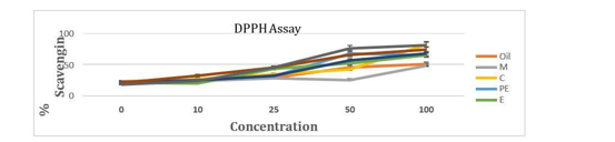
Figure 1: Antioxidant analysis by DPPH assay of oil and its extracts. The antioxidant activity of chloroform extract seems to be equivalent to NAC {N-acetyl cysteine, followed by aqueous extract, hexanoic extract and ethanoic extract showing nearly same potency, then petroleum ether extract is followed by our formulation, least in methanolic extract. The phytochemicals showed the highest antioxidant potency in semi-polar to non-polar solvents and the sesame oil is thereby used as a carrier, targeting the generation of oxidative stress at wound site.
FIGURE 2
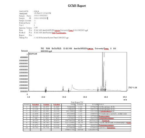
Figure 2: GCMS Report
FIGURE 3
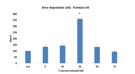
Figure 3: Cell proliferation assay (Dose Dependent) in Normal cell line. Cells were treated with varied concentration of oil. *p< 0.05
FIGURE 4
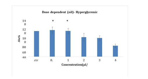
Figure 4: Cell proliferation assay (Dose Dependent) in Hyperglycemic cell line. Cells were treated with varied concentration of oil.*p< 0.05
FIGURE 5
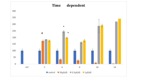
Figure 5: Time dependent cell proliferation assay in Normal cell- 30µl (Orange) and hyperglycemic cell were treated with 0.5µl (Grey) and 1µl (Yellow). #p< 0.1, *p< 0.05
FIGURE 6
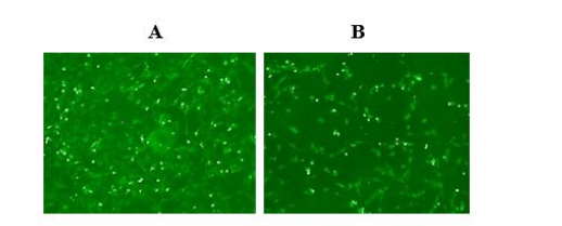
Figure 6: ROS generation in m5s cell. Cells were treated with various doses of Formulation for 2 hr and analysed by confocal and fluorescence microscopy. Representative images for (A) is Control and it gives green color fluorescence (B) Cells treated with 30µl oil. Cells with intracellular ROS are seen in less fluorescent. Control green colour compare to control. Intracellular ROS seen in more fluorescent compare to control. The green fluorescence represents stressful condition that remains almost similar in case of Control while in case of cells treated with oil, because of the potent antioxidant property, the ROS are scavenged. Hence the number of cell under stress is significantly less.
FIGURE 7
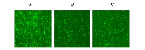
Figure 7: ROS generation in hyperglycaemic cells was (A) Control it gives green color fluorescence (B) and (C) Cells were treated with formulation (0.5µl, 1µl) showed less green fluorescence. (The green fluorescence represent stressful condition that remain almost similar in control while in case of cells treated with oil, because of the potent antioxidant property, the ROS are scavenged. Hence the number of cells under stress is significantly less
FIGURE 8
Figure 8: FACS performed in normal cells. A. The concentration of live cells in control is 95.03%, the cells undergoing pro-apoptosis are 3.73%. B. Cells treated with formulation are 94.7% showing similar result, the pro-apoptosis cell decrease to 3.17%
FIGURE 9
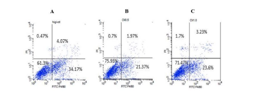
Figure 9: FACS performed in hyperglycaemic cells. (A) The concentration of live cells in control is 61.3%, cells treated with 0.5 µl (B) & 1 µl (C) formulation are 75.93% and 71.47% respectively. Under control condition the cells undergoing pro-apoptosis are 4.07%, while in 0.5 µl & 1 µl formulation treated cell decreases to 1.97% & 3.23% respectively
FIGURE 10
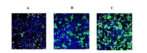
Figure 10: Phagocytosis activity in green fluorescence showed (A) control shows green fluorescence representing the phagocytic activity by macrophage. (B) and (C) Macrophage were treated with formulation showed significant phagocytic activity at 0.5µl and 1µl concentration
FIGURE 11
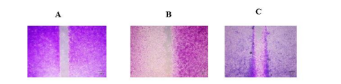
Figure 11: Cell migration assay (A) At 0 hr control, no migration was observed, (B) At 18hr control, cell migration elongation of dendrites in fibroblast and migration of border cells are indicative of invasiveness. (C) Formulation treated samples showed the significant migration of cells was observed
FIGURE 12
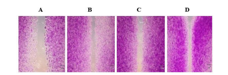
Figure 12: Cell Migration Assay in hyperglycaemic Cell (A) At 0 hr control, no migration was observed, (B) At 18hr control, cell migration elongation of dendrites in fibroblast and migration of border cells are indicative of invasiveness. (C) and (D) When treated with different concentrations of formulation i.e. 0.5µl and 1µl, gap filling started via cell migration
Tables at a glance
Figures at a glance