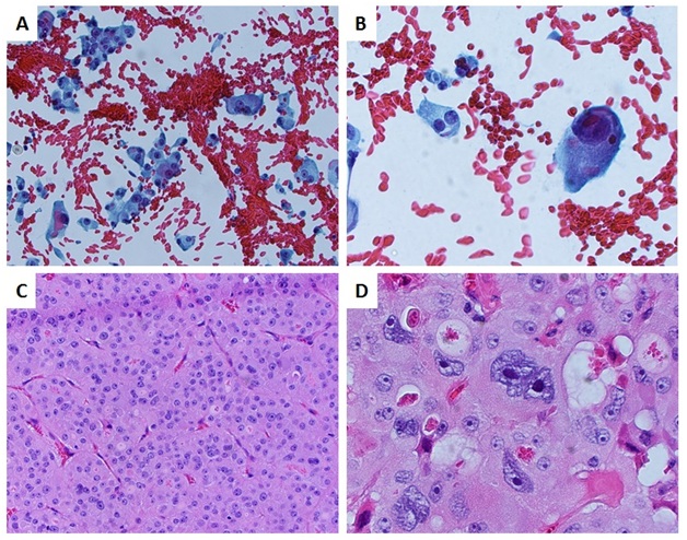
Figure 1 (A) Single cells, cell clusters and multinucleated giant cells exhibiting marked pleomorphism and anisonucleosis in a background of red blood cells (Papanicolaou stain, original magnification x200). (B) Single bizarre large cells present in a background of red blood cells (Papanicolaou stain, original magnification x400). (C) Small follicles of oncocytic cells that make up the bulk of the tumor (H&E stain, original magnification x200). (D) Small areas of oncocytic cells showing marked pleomorphism and nuclear atypia (H&E, original magnification x400).

Figure 2 (A) Sheets and single cells of an apparent Hürthle cell neoplasm in a background of red blood cells (Diff Quick stain, original magnification x100). (B) Single cells of an apparent Hürthle cell neoplasm in a background of red blood cells (Diff Quick stain, original magnification x200). (C) Anaplastic thyroid carcinoma with extensive necrosis, squamoid changes, and mitotic figures (H&E stain, original magnification x200) (D). Bizarre appearing cells with prominent enlarged nuclei, irregular nucleoli and multiple mitotic figures (H&E stain, original magnification x400).

Figure 3(A, Case 1) Clone of atypical cells negative for TTF-1 immunohistochemical staining; while background HCFC is positive (original magnification x200). (B, Case 1) Clone of atypical cells is BCL-2 negative; background HCC is positive (original magnification x200). (C, Case 1) Clone of atypical cells cyclin D1 is negative; with a background of positively staining HCFC (original magnification x200). (D, Case 2) The squamoid anaplastic cells are negative for TTF-1 staining; as compared with the background HCC (original magnification x200). (E, Case 2) The squamoid anaplastic cells are negative for BCL-2 staining, in comparison to the background HCC (original magnification x200). (F, Case 2) Cyclin D1 staining shows positive staining in the squamoid anaplastic cells; while negative in the Hürthle cells (original magnification x200).