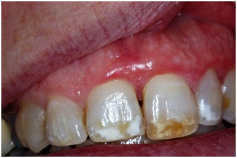
Figure 1 Case 1) Gingival erythema on facial gingiva of teeth #8, #9, biopsy result was “plasma cell gingivitis.” Image taken in December, 2008.
Figure 1 Case 1) Gingival erythema on facial gingiva of teeth #8, #9, biopsy result was “plasma cell gingivitis.” Image taken in December, 2008.
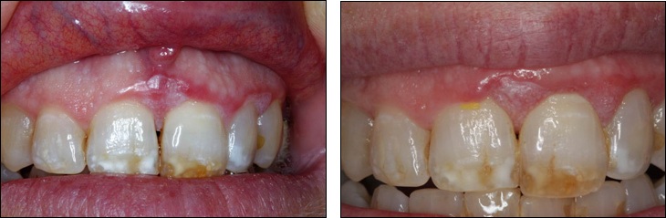
Figure 2 Case 1) Proliferative verrucous leukoplakia, the biopsy result was, “atypical papillary hyperplasia.” Image taken in June, 2013.
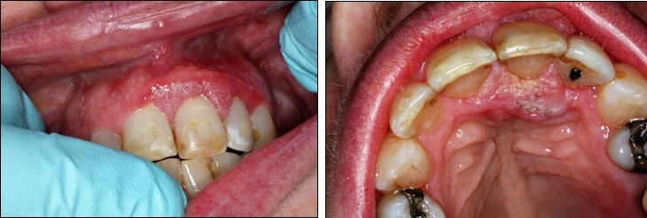
Figure 3Case 1) Recurrent lesion, pathology result was “atypical squamous proliferation, cannot exclude squamous cell carcinoma.” Image taken in December, 2013.
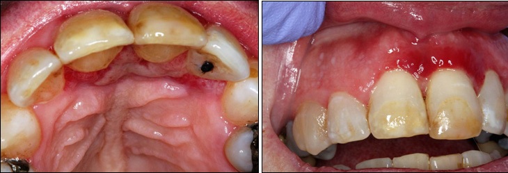
Figure 4Case 1) Post biopsy photos two weeks later. Image taken in January, 2014
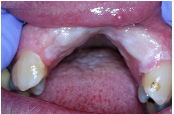
Figure 5Case 1) Post cancer treatment, including extraction of the involved teeth and bone resection. Image taken in 2016
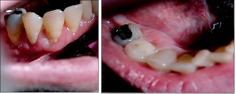
Figure 6Case 2) Erythema and inflammation, biopsy result was “consistent with lichen planus.” Image taken in 2004
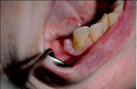
Figure 7Case 2) Recurrent lesion on distal of #27, the biopsy result was “consistent with lichen planus.” Image taken in 2005
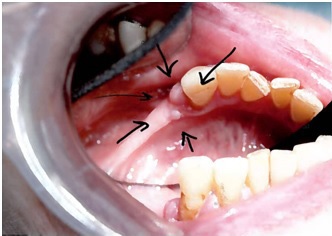
Figure 8Case 2) Recurrent lesion on distal of #27, the biopsy result was “epithelia hyperplasia and lichenoid gingivitis.” Image taken in 2011
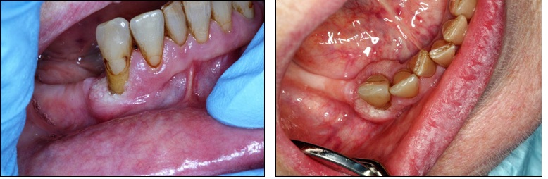
Figure 9Case 2) Proliferative Verrucous leukoplakia PVL on distal, buccal and lingual of #27, Image taken in 2013
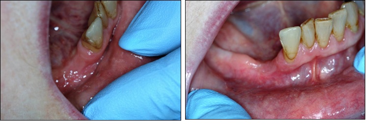
Figure 10Case 2) Complete removal of the lesion along with extraction of #27 was done, the area healed in two months