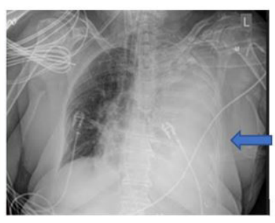
Figure 1 Anteroposterior chest x ray showing near complete opacification of left lung field (blue arrow)
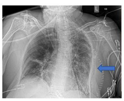
Figure 2 Anteroposterior chest x ray taken after SFTP removal. Without presence Of left lung field opacification ( Blue arrow)
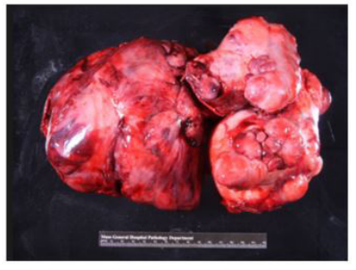
Figure 3 Gross pathological specimen showing large solitary pleural fibrous tumor
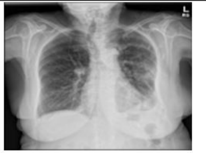
Figure 4 msteroanterior chest x ray taken at clinic follow up 2 weeks after surgery
Figures at a glance