
Atypical Presentation of a De Novo Pulmonary Embolism in a Young Female: A Case Report
Received Date: May 25, 2026 Accepted Date: June 04, 2026 Published Date: June 07, 2026
doi:10.17303/croa.2026.11.101
Citation: Huzaifa Usmani (2026) Atypical Presentation of a De Novo Pulmonary Embolism in a Young Female: A Case Report. Case Reports: Open Access 11: 1-4
Abstract
Pulmonary embolism (PE) is traditionally viewed as a secondary complication of deep vein thrombosis (DVT) originating in the lower extremities. However, "de novo" pulmonary embolism, thrombi forming directly within the pulmonary arterial vasculature, is increasingly recognized as a distinct clinical entity. Unlike traditional PE, de novo cases often occur in the absence of detectable peripheral venous clots and are frequently associated with localized inflammation, thoracic trauma, or chronic pulmonary conditions.PE categorized as "de novo" if comprehensive lower and upper extremity venous duplex ultrasound (VDUS) showed no evidence of DVT. Contrast to traditional pulmonary embolism (PE), where thrombi usually migrate from large deep veins in the legs, de novo pulmonary embolism is characterized by in situ thrombosis. This means the clot forms directly within the pulmonary arterial tree. The primary sites for de novo pulmonary embolism include, Segmental and Subsegmental Arteries, because de novo PE is frequently driven by localized inflammation or "immunothrombosis," the clots tend to form in these smaller, peripheral vessels rather than the larger central arteries. Thrombi often develop in vessels adjacent to lung pathology, such as areas affected by, severe pneumonia, acute Respiratory Distress Syndrome, Chronic Obstructive Pulmonary Disease exacerbations. Vessels Adjacent to Thoracic Trauma, in cases of blunt chest trauma or lung contusion, the site of the clot is typically the vasculature directly involved in or immediately surrounding the injured lung tissue.
Keywords: Pulmonary Embolism; Deep Vein Thrombosis; Thoracic Trauma; Chest Trauma; Pneumonia; Lung Contusion
Introduction
Pulmonary Embolism (PE) is an acute serious condition that could be life threatening if not diagnosed and treated promptly. The pathology arises in lung artery which is blocked by some substance that travelled from elsewhere in the body to lung arteries, mainly blood clot from leg or pelvis.PE is life threatening and dramatic complication of DVT. PE and DVT are the manifestation of same cause namely Thrombo-Embolic disease. The most common form of emboli that causes PE is blood clot from lower limb and pelvis.DE NOVO PULMONARY EMBOLISM (DNPE) refers to PE that occurs without evidence of a Deep Vein Thrombosis. Unlike classic PE where a clot travel from leg, DNPE is thought to result from localized lung inflammation, endothelial damage or high level pro coagulant factor following severe injury or illness. Other condition that causes Pulmonary Embolism are, amniotic emboli during child birth, tumor emboli from a malignant disease, fat emboli originating from bone or bone marrow in patient of fracture or trauma. Studies show widely varying rates of PE without DVT, ranging from 11% to 59%.
Case Report
A 31-year-old female came to ER with history of pain and cramps in her right leg an hour ago at work. The patient denies any chest pain, shortness of breath, palpitation, sweating or productive cough. Patient denies any history of travel, recent surgery or immobilization. Patient was on oral contraceptive pills previously but had discontinued using OCP in recent times. On examination there is mild calf tenderness but no swelling. There is no shortness of breath or dyspnea. Her chest and heart examination were normal. No tachycardia or tachypnea noted. Her vitals read as B.P-112/70mmhg, HR-75/min, RR-18cpm, and SPO2-98% on room air. ECG showed normal sinus rhythm. Echocardiography reveals no abnormal finding.
Labs and U/S DVT was done. D-dimer was high 6.30(range 0-0.5), Troponin was negative, rest of the labs including inflammatory markers were all normal. U/S DVT reveals no clot in leg vein. Patient in view of high D-dimer was scanned for lung vessels, CT-pulmonary angiogram, revealed multiple contrast filling defects in right interlobar artery, right lower lobe posterior and lateral segmental arteries, left lingular artery, bifurcation of the left lower lobe posterior and lateral segmental arteries, left lower lobe posterior segmental arteries,suggesting of multilple pulmonary emboli. Patient was given enoxaparin and admitted. Patient remained stable for the next 24 hrs and was discharged on anticoagulant.
Conclusion
The clinical characterization of de novo pulmonary embolism, arising directly within the pulmonary vasculature rather than embolizing from deep vein thrombosis, marks a significant shift in our understanding of venous thromboembolism. Mounting evidence highlights distinct pathophysiological pathways for de novo events, often driven by localized pulmonary inflammation, endothelial dysfunction, and regional hypoxia, rather than traditional systemic hypercoagulable states. Recognizing these mechanistic differences has profound implications for patient management. While conventional PE guidelines heavily rely on prolonged anticoagulation aimed at preventing recurrent systemic emboli, de novo PE may require a more tailored approach. Future strategies should prioritize targeted therapies that address underlying pulmonary inflammation and localized vascular health, rather than relying solely on standard systemic clot prevention.Ultimately, distinguishing de novo PE from traditional thromboembolic disease is essential for refining risk stratification, optimizing therapeutic interventions, and improving long-term patient outcomes. Continued clinical research and molecular studies are imperative to fully elucidate these localized pathways and establish dedicated diagnostic and treatment protocols for this distinct patient population.
Pulmonary Embolism can be a difficult diagnosis that can be missed because of non-specific symptoms presentation in emergency, as was the case in this patient as she was symptoms free. This patient was typical case of De Novo PE with atypical presentation and high suspicious of index is required for diagnosis. This patient have low risk factor of developing PE as only risk factor was history of OCP consumption, as OCP increased the risk of production of clotting factor (procoagulant) and decreased natural anticoagulant by acquired resistance to activated PROTEIN C . A physician good clinical eye and keeping low threshold for such patient can be helpful in establishing the diagnosis, as an early diagnosis is fundamental for better prognosis and immediate treatment is highly effective.
- Van Gent et al. (2014) Pulmonary embolism without deep venous thrombosis: de novo or missed deep venous thrombosis.
- Wongweerakit et al. (2025) De novo pulmonary embolism following chest trauma: fact or fiction.
- Leibowitz et al. Challenging Traditional Paradigms in Posttraumatic Pulmonary Embolism.
- Zinoune et al. (Immediate Pulmonary Embolism / Case Reports).
- Annals of Vascular Surgery (2012) Pulmonary Embolism without Deep Venous Thrombosis.
FIGURE 1
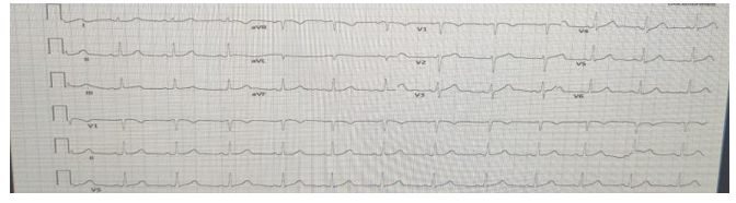
Figure 1
FIGURE 2
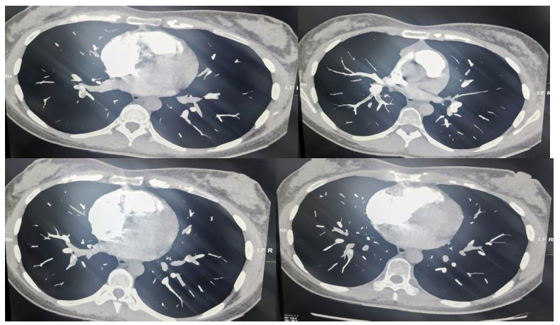
Figure 2
Figures at a glance