
Stroke and Posterior Medullopathy in a Patient with Suspected Neuroborreliosis: Case Report
Received Date: July 21, 2026 Accepted Date: August 08, 2026 Published Date: August 11, 2026
doi:10.17303/croa.2026.11.103
Citation: Sima Ataollahi Eshkoor, Abdul Mojib Sarwari, Masoud Falah, Jolanta Gaudesiene, Plamen Anzhelov Bekyarov (2026) Stroke and Posterior Medullopathy in a Patient with Suspected Neuroborreliosis: Case Report. Case Reports: Open Access 11: 1-10
Abstract
A 50-year-old man was admitted with recurrent falls due to progressive weakness in the left lower extremity. Initial neuroimaging MRI revealed a subacute infarction in the pons and a symmetric posterior medullopathy in the cervical spinal cord in the form of inverted V shape suggestive subacute combined degeneration. Subsequent cerebrospinal fluid (CSF) analysis showed mononuclear pleocytosis and elevated Borrelia IgG, which resulted in the diagnosis of neuroborreliosis and the need for appropriate treatment. It is concluded that the diagnostic complexity of concurrent pontine infarction and inflammatory medullopathy in patients with multifocal neurological symptoms requires extensive clinical insight, integrating radiological and microbiological findings.
Keywords: Lyme, Myelopathy, Neuroborreliosis, Stroke
Introduction
Lyme borreliosis (LB), also known as Lyme disease, is a multisystem, multistage zoonotic infection caused by spirochetes of the Borrelia burgdorferi sensu lato complex, transmitted through the bite of infected Ixodes ticks [1]. It is the most common vectorborne disease in the northern hemisphere [2,3]. The clinical presentation of Lyme borreliosis is variable and typically progresses through three overlapping stages known as early localized, early disseminated, and late disseminated infection.
The disease often begins with erythema migrans, a characteristic skin lesion appearing days to weeks after the tick bite. In later stages, the infection may disseminate, leading to neurological, cardiac, dermatological, or articular involvement, which can occur months to years after the initial infection. In the late disseminated stage, Lyme disease may manifest as chronic arthritis or persistent neurological symptoms, including encephalopathy, cognitive disturbances, or peripheral neuropathy, often accompanied by spinal radicular pain, distal paresthesia, or hypoesthesia [2]. Involvement of the central or peripheral nervous system occurs in Lyme neuroborreliosis (LNB) that happens in approximately 10–15% of Lyme disease cases and can affect any part of the nervous system [1]. The most frequent presentations are lymphocytic meningitis, radiculoneuritis, and cranial neuritis that can happen separately or in combination [4]. Severe forms, such as stroke, cerebral vasculitis [1,5], encephalitis, or myelitis, may also occur, although rarely [5].
Patient History
A 50-year-old man with known obstructive sleep apnea and asthma was admitted to the emergency ward after two falls, followed by progressively increasing weakness on his left side of body. He had experienced a growing inability to walk independently. In addition, for approximately one and a half months he had developed neck pain and a tightening sensation in both shoulders, as well as tingling in both upper extremities and coordination difficulties in his left arm. He reported no sensory loss but noted unsteadiness and described his gait as similar to that of a drunk person. He also reported constant night sweats during the summer months and an unintentional weight loss of approximately 15 kg over 3 months. He denied tick bites, recent travel outside Europe, or high-risk sexual behavior, as well as heavy alcohol or cannabis use. His family history was notable for the death of his mother from a cerebral tumor at age 51.
Clinical Findings
On admission, the patient was alert, oriented, and afebrile (T 36.5 °C). Vital signs were stable with blood pressure 140/90 mmHg, heart rate 77 bpm, and oxygen saturation 100%. Neurological examination revealed mild left facial paresis, tongue deviation to the left, and mild dysarthria. In addition, there were found severe left-sided lower limb paresis and mild left upper limb paresis with preserved tone and reflexes as well as the presence of left-sided Babinski sign. Sensation was intact to light touch and pinprick in all extremities. There were also found impaired coordination due to weakness, gait ataxia requiring assistance, and extensor plantar response on the left side.
Laboratory Findings
Routine blood tests were within normal limits, including hemoglobin (8.8 mmol/L), electrolytes, renal and liver function tests, CRP (<1 mg/L), and vitamin B12 levels. Lumbar puncture revealed a mononuclear pleocytosis of 74 cells/µL, mildly elevated lactate (2.7 mmol/L), normal glucose (2.3 mmol/L), and markedly elevated CSF IgG (790 mg/L). CSF CXCL13 levels were also elevated (250–500 pg/mL), and oligoclonal immunoglobulin bands were present. The patient had a positive Borrelia-specific intrathecal antibody index (Borrelia IT), supporting intrathecal production of Borrelia-specific antibodies. HIV and syphilis serology were negative.
The combination of a compatible neurological presentation, inflammatory CSF findings with mononuclear pleocytosis, elevated CSF IgG and CXCL13, and a positive Borrelia-specific intrathecal antibody index supported the diagnosis of Lyme neuroborreliosis. In particular, the positive intrathecal Borrelia antibody response provided evidence of a Borrelia-specific immune response within the central nervous system (CNS), while the CSF pleocytosis and elevated CXCL13 supported an active inflammatory process. The presence of oligoclonal bands further indicated intrathecal immunoglobulin production, although these findings are not specific for neuroborreliosis. Taken together, the clinical and paraclinical findings were considered consistent with neuroborreliosis with multifocal CNS involvement.
Neuroimaging
MRI Brain (19/09/2025)
FLAIR and diffusion-weighted imaging revealed subacute infarctions in the central and right pontine regions without evidence of hemorrhage, mass lesion, or meningeal enhancement (Fig 1).
MRI Cervical Spine (19/09/2025)
Symmetric bilateral hyperintensity in the dorsal columna from C2–C6 on T2-weighted sequences forming a “V-shaped” pattern, consistent with posterior medullopathy. No evidence of compressive myelopathy or contrast enhancement. Degenerative changes were mild (Fig 2-4). These findings were initially interpreted as subacute combined degeneration of the spinal cord, though serum B12 was normal.
Hospital Course and Management
Neurological examinations and neuroradiological consultations were obtained, and several differential diagnoses were considered because of the combination of progressive multifocal neurological deficits, posterior column abnormalities on spinal MRI, pontine infarction, and systemic symptoms including substantial weight loss, night sweats, and lymphadenopathy.
The symmetric posterior column signal abnormality initially raised the possibility of subacute combined degeneration of the spinal cord. However, this diagnosis was considered unlikely because the patient's serum vitamin B12 level was normal and the presence of a mononuclear CSF pleocytosis was not consistent with an isolated nutritional myelopathy. Nitrous oxide-induced myelopathy was also considered in the differential diagnosis, but the patient denied relevant substance use or other reported risk factors. Autoimmune and paraneoplastic inflammatory myelitis were considered because of the multifocal neurological involvement and systemic symptoms. HIV and syphilis serology were negative. A CT scan of the thorax and abdomen was performed as part of the malignancy work-up in view of the patient's weight loss, night sweats, and lymphadenopathy, and no alternative cause was identified. Further investigations did not support neurosarcoidosis.
Because of the atypical combination of a pontine infarction and inflammatory posterior medullopathy, neuroborreliosis with possible vasculitic involvement was considered. The diagnosis was supported by the inflammatory CSF profile, including mononuclear pleocytosis, elevated CSF IgG and CXCL13, and, importantly, a positive Borrelia-specific intrathecal antibody index. The absence of radiological findings suggestive of multiple sclerosis (MS) or neuromyelitis optica spectrum disorder (NMOSD) on subsequent MRI of the neuraxis further reduced the likelihood of these alternative inflammatory demyelinating disorders.
Empirical treatment with intravenous ceftriaxone (2 g daily) was initiated for suspected neuroborreliosis. The patient subsequently received a 14-day course of intravenous ceftriaxone in the Department of Infectious Diseases. Following completion of antimicrobial treatment, he was transferred to the Department of Neurology for further neurological assessment and rehabilitation. During follow-up, he remained hemodynamically stable, with gradual improvement in activities of daily living but persistent spasticity of the left upper extremity. Following rehabilitation, he was referred for botulinum toxin injections.
Diagnosis and Treatment
The diagnosis of neuroborreliosis was based on the combination of the patient's progressive neurological symptoms and objective evidence of CNS inflammation and Borrelia-specific intrathecal immune activation. CSF analysis demonstrated a mononuclear pleocytosis of 74 cells/µL and elevated CSF IgG (790 mg/L), indicating an inflammatory process within the CNS. The markedly elevated CSF CXCL13 level (250–500 pg/mL) provided additional supportive evidence of intrathecal neuroinflammation and B-cell recruitment.
Most importantly, the positive Borrelia-specific intrathecal antibody index (Borrelia IT) supported intrathecal production of Borrelia-specific antibodies and therefore provided specific evidence supporting neuroborreliosis in the context of the patient's clinical presentation and inflammatory CSF findings. The presence of oligoclonal bands indicated intrathecal immunoglobulin production but was considered nonspecific.
The patient was treated with intravenous ceftriaxone for 14 days. Following antimicrobial therapy, he was transferred to the Department of Neurology for further assessment and multidisciplinary rehabilitation. MRI of the neuraxis was subsequently performed to investigate alternative inflammatory demyelinating disorders, including MS and NMOSD, but showed no radiological findings suggestive of either condition. Further paraclinical investigations did not support neurosarcoidosis or syphilis. Despite antimicrobial treatment and rehabilitation, the patient had persistent spasticity of the left upper extremity and was subsequently referred for botulinum toxin treatment.
Discussion
There are various manifestations of LNB, including aseptic meningitis, erythema migrans rashes, Bannwarth’s syndrome [6], myelitis [1,7,8], cerebral vasculitis [1,9], and stroke [6,9] that present differently in American and European patients [6].
These differences in the manifestations of LNB between American and European patients, have been attributed to genetic variations among the Borrelia strains present in each region [6]. It has been reported that B. burgdorferi sensu stricto is isolated from the CSF of American patients, whereas B. garinii or B. afzelii are more commonly isolated from European patients [10]. Moreover, it could also be due to a stronger inflammatory response in the nervous system, more resistance to the host’s immune defenses, and probably more persistence in neural tissues in European patients [6].
American CNS Lyme patients typically present with aseptic meningitis and often with erythema migrans rashes, whereas European CNS Lyme more commonly presents as a painful polyradiculitis known as Bannwarth’s syndrome and only rarely with erythema migrans rashes [6]. In addition to myelitis, cerebral vasculitis and stroke have been reported in some cases of European patients [9]. Various reports [9,11] have indicated stroke as a complication of Lyme disease especially in European patients, yet it is rarer in American patients. The occurrence of Stroke in European patients is probably due to Lyme meningovasculitis or reversible cerebral vasoconstriction syndrome probably mediated by an inflammation of intracranial vasculature with subsequent narrowing and decreased blood flow [6].
Stroke has been reported after non-specific complaints, such as headache, fatigue, difficulty concentrating, nausea and vomiting as well as after erythema migrans, reported in only two cases, including the one American patient [6]. It has reported that stroke occurs sometimes after Lyme antibiotic treatment [12]. As it is a rare complication of disease, therefore there is no need to screen Lyme serology in all patients with acute stroke [6].
Vasculopathy originating from direct infiltration of the vessel wall by spirochaetes, spread of inflammation from adjacent meninges and/or from deposition of immune complexes, remains still unclear [6]. It has been reported that the impact of inflammation on the arteries can sometimes be permanent. It sounds also, the inflammation occurred due to this infection probably involves arterial segments that are already affected by other processes, such as atherosclerosis [6]. Despite permanent inflammation and clinically affection due to Lyme infection repeating antibiotic therapy after Lyme infection is not yet recommended [6]. The rare and sever complications of LNB infection are encephalitis, myelitis [1,8] and cerebral vasculitis [1].
Myelitis happens often in European LNB. As instance, it has been reported in approximately 7% of Lyme patients in Germany diagnosed by MRI spine imaging [1,13,14].
It most often presents in the form of radiculomyelitis [15], but it can also be seen as transverse myelitis (ATM) [8,16], longitudinally extensive myelitis (LETM) [17-19], meningomyelitis or meningoradiculomyelitis [20,21], posterior column–predominant myelitis (Radzišauskienė et al., 2023), and gray matter–predominant myelitis. The gray matter–predominant form is extremely rare [22]. This our case demonstrates a rare overlap of ischemic stroke and inflammatory spinal cord involvement. The coexistence of a pontine infarct and posterior medullopathy raises the possibility of a systemic process affecting both the central and peripheral nervous systems. Furthermore, MRI of the cervical spinal cord revealed symmetric bilateral hyperintense signals in the dorsal columns, which is known as inverted V-shaped sign and a characteristic radiological feature of vitamin B12 deficiency myelopathy [23-25].
Although MRI findings were suggestive of subacute combined degeneration, normal B12 levels and presence of CSF mononuclear pleocytosis argue against nutritional deficiency. Elevated Borrelia IgG and CXCL13 supported an inflammatory or infectious etiology, possibly neuroborreliosis with multifocal CNS involvement. In addition, a pontine infarction was identified in this patient. Although pontine infarction due to vasculitic mechanisms in neuroborreliosis is rarely reported, it is biologically plausible [26]. Malignancy and chronic infection were considered in the differential diagnosis due to the presence of systemic symptoms, including weight loss, night sweats, and lymphadenopathy, but were subsequently excluded following appropriate diagnostic evaluation.
Conclusion
We report an unusual case of subacute pontine infarction coexisting with posterior medullopathy and inflammatory CSF findings in a middle-aged man. The combination of multifocal neurological deficits, a posterior column-predominant spinal cord lesion, pontine infarction, CSF mononuclear pleocytosis, and evidence of intrathecal Borrelia-specific antibody production illustrates the diagnostic complexity of neuroborreliosis with multifocal CNS involvement. The key clinical lesson is that posterior column abnormalities on spinal MRI should not automatically be attributed to vitamin B12 deficiency, particularly when the clinical presentation is atypical or accompanied by inflammatory CSF findings. In patients with combined brainstem and spinal cord lesions, infectious etiologies such as neuroborreliosis should be considered alongside inflammatory, nutritional, autoimmune, paraneoplastic, and vascular causes. A comprehensive diagnostic evaluation incorporating clinical findings, CSF analysis, Borrelia-specific intrathecal antibody testing, and appropriate investigation of alternative diagnoses can help establish the underlying cause and guide timely antimicrobial treatment. This case emphasizes the importance of maintaining a broad differential diagnosis in patients with atypical multifocal neurological presentations, as early recognition and treatment of infectious causes may prevent further neurological deterioration.
Notes on Patient Consent
Patient consent for publication of the clinical information and results presented in this article was obtained.
Funding Information
This work received no specific grant from any funding agency.
Conflicts of Interest
The authors declare that there are no conflicts of interest.
- Garkowski A, Łebkowska U, Kubas B, Garkowska E, Rutka K, et al. (2020) Imaging of Lyme neuroborreliosis: A pictorial review. Paper presented at the Open forum infectious Diseases.
- Kullberg BJ, Vrijmoeth HD, van de Schoor F, Hovius JW (2020) Lyme borreliosis: diagnosis and management. bmj, 369.
- Steere AC, Strle F, Wormser GP, Hu LT, Branda JA, Hovius JW, et al. (2016) Lyme borreliosis. Nature reviews Disease primers, 2: 1-19.
- Ford L, Tufts DM (2021) Lyme neuroborreliosis: Mechanisms of B. burgdorferi infection of the nervous system. Brain sciences, 11: 789.
- Zajkowska J, Garkowski A, Moniuszko A, Czupryna P, Ptaszyńska-Sarosiek I, Tarasów E, et al. (2015) Vasculitis and stroke due to Lyme neuroborreliosis–a review. Infectious diseases, 47: 1-6.
- Li S, Vytopil M, Hreib K, Craven DE (2015) Lyme disease presenting as multiple ischaemic strokes. Practical Neurology, 15: 284-8.
- Popli K, Khamishon R, Nutakki A, Probasco J (2024) Acute Transverse Myelitis Due to Neuroborreliosis: A Case Report (P3-13.007). Paper presented at the Neurology.
- Radzišauskienė D, Urbonienė J, Jasionis A, Klimašauskienė A, Malickaitė R, Petrulionienė A, et al. (2023). Clinical and epidemiological features of Lyme neuroborreliosis in adults and factors associated with polyradiculitis, facial palsy and encephalitis or myelitis. Scientific Reports, 13: 19881.
- Topakian R, Stieglbauer K, Nussbaumer K, Aichner FT (2008) Cerebral Vasculitis and Stroke in Lyme NeuroborreliosisTwo Case Reports and Review of Current Knowledge. Cerebrovascular Diseases, 26: 455-61.
- Pachner AR, Steiner I (2007) Lyme neuroborreliosis: infection, immunity, and inflammation. The Lancet Neurology, 6: 544-52.
- Hammers-Berggren S, Gröndahl A, Karlsson M, Von Arbin M, Carlsson A, Stiernstedt G (1993) Screening for neuroborreliosis in patients with stroke. Stroke, 24: 1393-6.
- Reik Jr L (1993) Stroke due to Lyme disease. Neurology, 43: 2705.
- Sáenz JA, de la Gala DH, García AA, Venero CS, de Lucas EM (2021) Atypical bacterial infections of the central nervous system transmitted by ticks: An unknown threat. Radiología (English Edition), 63: 425-35.
- Schwenkenbecher P, Pul R, Wurster U, Conzen J, Pars K, Hartmann H, et al. (2017) Common and uncommon neurological manifestations of neuroborreliosis leading to hospitalization. BMC infectious diseases, 17: 90.
- Opielka M, Opielka W, Sobocki BK, Starzynska A (2020) Subacute transverse myelitis with optic symptoms in neuroborreliosis: a case report. BMC neurology, 20: 244.
- Dumic I, Vitorovic D, Spritzer S, Sviggum E, Patel J, Ramanan P (2019) Acute transverse myelitis–A rare clinical manifestation of Lyme neuroborreliosis. IDCases, 15: e00479.
- Abbas SA, Costa J, Saba J, Matta C, Abboud H (2025) Longitudinally extensive transverse myelitis: Etiologies and common diagnostic pitfalls. Neuroimmunology Reports, 7: 100255.
- Contentti EC, Hryb J, Leguizamón F, Di Pace J, Celso J, Knorre E, Perassolo M (2017) Differential diagnosis and prognosis for longitudinally extensive myelitis in Buenos Aires, Argentina. Neurología (English Edition), 32: 99-105.
- Nightingale H, Witherick J, Wilkins A (2011) Diagnosis of longitudinally extensive transverse myelitis. Case Reports, 2011: bcr1020103444.
- Gómez-Eguílaz M, Gómez-Cerquera J, Calvo-Pérez L, Oteo J (2014) Neuroborreliosis: A single-hospital series of 7 cases. Neurologia (Barcelona, Spain), 31: 137-9.
- Riggle C, Brissette CA (2021) Implications and Aspects of Lyme Neuroborreliosis. ECCMID, 72.
- Lindland ES, Solheim AM, Andreassen S, Bugge R, Eikeland R, Reiso H, et al. (2023). Dynamic contrast-enhanced MRI shows altered blood–brain barrier function of deep gray matter structures in neuroborreliosis: a case–control study. European Radiology Experimental, 7: 52.
- Narra R, Mandapalli A, Jukuri N, Guddanti P (2015) “Inverted V sign” in sub-acute combined degeneration of cord. Journal of Clinical and Diagnostic Research: JCDR, 9: TJ01.
- Nouri A, Patel K, Montejo J, Nasser R, Gimbel DA, Sciubba DM, et al. (2019) The role of vitamin B12 in the management and optimization of treatment in patients with degenerative cervical myelopathy. Global Spine Journal, 9: 331-7.
- Stabler SP (2013) Vitamin B12 deficiency. New England Journal of Medicine, 368: 149-60.
- Van Snick S, Duprez T, Kabamba B, Van De Wyngaert F, Sindic C (2008) Acute ischaemic pontine stroke revealing lyme neuroborreliosis in a young adult. Acta neurologica belgica, 108: 103-6.
FIGURE 1
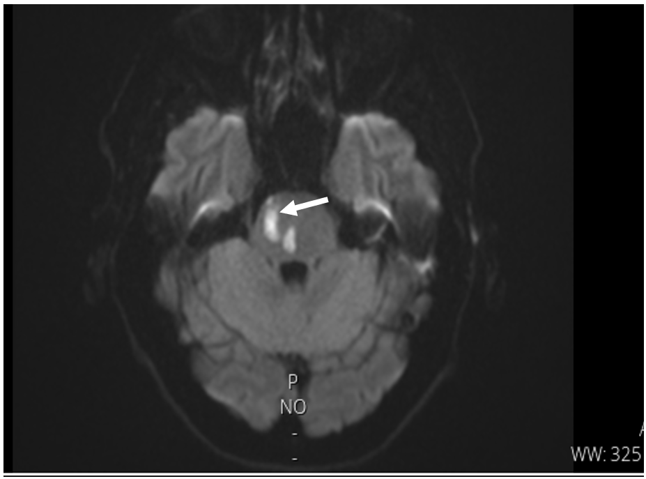
Figure 1: Pontine stroke at the patient with neuroborreliosis
FIGURE 2
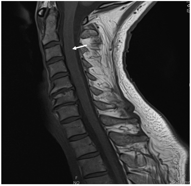
Figure 2: Myelopathy induced in Neuroborreliosis (Sagittal T1W image)
FIGURE 3
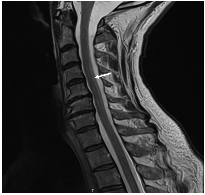
Figure 3: Myelopathy induced in Neuroborreliosis (Sagittal T2W image).
FIGURE 4
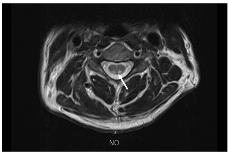
Figure 4: Myelopathy induced in Neuroborreliosis (Axial T2W image).
Tables at a glance
Figures at a glance