
Restoration of Severely Atrophic Maxilla Using Extra-Sinus Zygomatic Implants Without Bone Augmentation: A Case Series from Our Clinical Experience
Received Date: August 21, 2026 Accepted Date: October 02, 2026 Published Date: October 06, 2026
doi:10.17303/croa.2026.11.104
Citation: Büşra Nida Üzümcü DDS, Deniz Gölpek DDS, Arda Kement DDS, Fatma Nur Çalap İçli DDS, Serhat Yalçın DDS (2026) Restoration of Severely Atrophic Maxilla Using Extra-Sinus Zygomatic Implants Without Bone Augmentation: A Case Series from Our Clinical Experience. Case Reports: Open Access 11: 1-9
Abstract
Objective: To evaluate the success of zygomatic implants placed using the extra-sinus technique combined with endosseous dental implants for the restoration of the severely atrophic maxilla, aiming to avoid the disadvantages of augmentation methods.
Materials and Methods: In 12 patients with severely atrophic maxilla, a total of 29 zygomatic implants were placed, passing through the lateral wall of the maxillary sinus without any augmentation methods. These implants were distributed as 4 implants in 4 patients, 2 implants in 5 patients, and 1 implant in 3 patients.
Results: No intraoperative or postoperative complications were observed, and all implants successfully achieved osteointegration. The prosthetic rehabilitation of the patients was successfully completed.
Conclusion: The combination of zygomatic implants passing through the lateral wall of the maxillary sinus with endosseous dental implants provided a successful aesthetic and functional treatment alternative to existing augmentation methods for the severely atrophic maxilla. Predictable outcomes were achieved through careful preparation of the surgical area, appropriate technique selection based on clinical and radiological evaluations, and the surgeon's experience.
Keywords: Endosseous Dental Implant, Maxillary Resorption, Zygomatic Implant, Augmentation
Introduction
"Severe horizontal and vertical bone losses in the maxilla can be caused by various systemic and local factors such as early tooth loss, periodontal diseases, or tumor resection (1). Treatment options for the rehabilitation of these conditions include conventional total prostheses, implant-supported total prostheses using bone grafts, customized new-generation subperiosteal implants, and zygomatic implants (2). Zygomatic implants are a frequently used method for the treatment of the severely atrophic maxilla. Regarding the study design and clinical methodology of this implants, the selection process follows the strict clinical pathways outlined in current implant dentistry consensus statements, which advocate for systematic patient selection to ensure predictable long-term survival rates (23). According to the ITI consensus guidelines, the primary patient selection criteria for zygomatic implant interventions mandate adult individuals presenting with severe maxillary atrophy (Cawood and Howell Class V or VI) or extensive bone loss due to trauma or pathology, specifically where conventional block grafting or sinus augmentations are contraindicated or declined by the patient (23). Conversely, candidates with active sinonasal infections, uncontrolled systemic conditions, or recent craniofacial radiation therapy are excluded to mitigate biological complications (23).
Zigomatic implants interventions involve four different application techniques: intrasinus, sinus slot, extra-sinus, and extramaxillary (2,3). In the selection of the surgical technique, mechanical durability against masticatory forces and aesthetic success are important factors (4). The extra-sinus approach is preferred in patients with pronounced buccal concavity, where the neck of the zygomatic implant is positioned close to the alveolar crest (5). Implants are generally performed under general anesthesia for patient comfort and ease of surgical operation, although some cases report that it can also be performed under local anesthesia and sedation (6-8). The literature reports a success rate of 82-100% for zygomatic implants, making them a predictable treatment option (1,9).
Materials and Methods
From October 2017 to January 2024, 12 patients with severely atrophic maxilla presented to Istanbul Kent University and Dentram Dental Clinic (Levent-Bağdat, Istanbul). The study included 7 female and 5 male patients aged 36-65, all with periodontal disease, most of whom were edentulous. The patients were generally in good health, with 3 having hypertension and 5 diabetes, all under control. The surgeries were performed under general anesthesia. This study was conducted in accordance with the ethical principles of the Declaration of Helsinki as revised in 2013. During the procedur ensuring that comprehensive written informed consent was explicitly obtained from each patient prior to the initiation of any clinical procedures.
Surgical Procedure (Figures 1-6)
Clinical and radiological examination of 12 cases revealed that according to the Bedrossian Classification for the maxilla, 8 cases had sufficient bone in only Zone 1 for endosseous dental implants. The Quad Zygoma procedure was applied to the remaining 4 cases.
Four weeks before the surgery, patients' 25-hydroxy vitamin D levels were checked. It was found that patients' 25-hydroxy vitamin D levels were in the range of 17-28 ng/ml, deemed insufficient for osteointegration. Oral vitamin D drops were given 3 weeks before the surgery. Sedative medications were administered 1 hour before the operation. Fiber-optic nasotracheal intubation and total muscle relaxant were applied under general anesthesia. At the beginning of the operation, an intravenous dose of steroids was given. Local anesthesia was applied to the buccal and palatal regions of the maxilla. Full-thickness horizontal and vertical incisions were made along the crest line close to the palate with a 15c scalpel, and dissection of the periosteum and soft tissues along the zygomatic notch towards the zygomatic bone was performed. The lateral surface of the maxilla was clearly exposed. Before implant placement, infraorbital foramina and zygomatic buttresses were identified for anatomical orientation. The palatal flap was tied with silk sutures to increase the field of view (Figure 2). After using a round bur, implant sites were prepared using pilot and final burs. To facilitate implant placement, the apical part of the implants was directed towards the zygomatic bone using an angled drill. The remaining part of the implants was inserted into the implant site using a manual carrier, ensuring proper orientation of the implant heads. No augmentation method was used (Figure 2).
The surgical field was sutured with 6.0 polyglycolide rapid suture. Painkillers, antibiotics, steroids, gastric protectors, and mouthwashes were prescribed. Using the mentioned technique, a total of 29 zygomatic implants were placed in the first-second premolar regions of the upper (left and/or right) maxilla. In 3 cases, the zygomatic implant was solitary and located on only one side of the upper maxilla. Additionally, a total of 36 endosseous dental implants were used to restore the upper jaw in 9 patients. Generally, NobelZygoma implants (available in 30, 32.5, 35, 37.5, 40, 42.5, 45, 47.5, 50, and 52.5 mm lengths) and NobelParallel™ Conical Connection implants were selected in dimensions suitable for the patients' specific anatomical configurations. Temporary prostheses were delivered on the same day as the surgery, significantly improving the patients' quality of life. All patients agreed to adhere to the oral hygiene protocol and recall appointments. The postoperative period was uneventful, and there was no fibrotic soft tissue growth that would prevent bone healing. All implants successfully achieved osteointegration (Figure 3) and no postoperative complications such as sinusitis or mucositis were observed within 1 week.
Healing abutments were placed following soft tissue healing (Figure 4).
Prosthetic restorations were successfully completed within 3 months without any adverse events reported by the patients (Figures 5, 6, 7). At 12-month follow-up, no implant loss was observed.
Discussion
Patients with minimal vertical and horizontal bone volume require advanced surgical reconstruction techniques to restore chewing and speech functions (10). Zygomatic implants were first introduced in the late 1990s by Dr. Per Ingvar Branemark. Although relatively new among treatment options for the atrophic maxilla, zygomatic implants have evolved from the remote anchorage implant concept developed by Parel and colleagues (11,12). Recent clinical case reports indicate that the use of zygomatic implants in the treatment of atrophic maxilla, by eliminating the need for bone grafts used in other advanced surgical treatments, shortens the expected treatment time for the maturation of these bone grafts and reduces the healing time of the implants, achieving long-term successful results (11,13).
A recent systematic review based on 68 studies involving 4556 zygomatic implants in 2161 patients reported a 12-year survival rate of 95.21%. 14 Brånemark et al., in their study on 28 patients, conducted regular clinical and radiographic examinations of 56 zygomatic implants and reported that zygomatic implants showed a 10-year survival rate of 94.3%, indicating that they are a predictable treatment option (15). Despite the high success rate of zygomatic implants, complications such as sinusitis, peri-implant soft tissue infections, nerve paresthesia, oroantral fistula, zygoma fractures, and orbital complications have been reported (15-18).
Various treatment methods have been applied for the oral rehabilitation of patients with severely atrophic edentulous maxilla (19,20). Among these treatment methods, zygomatic implants are often preferred over bone augmentation methods, which are typically performed in two stages, due to lower costs, time savings from the single-stage application of zygomatic implants, and their high success rate (21,22). However, because zygomatic implant procedures are major surgeries and are adjacent to critical structures such as the infraorbital nerve and the orbit, they require extensive experience. The most common complications in zygomatic implants include infraorbital or zygomatic facial nerve paresthesia, sinusitis, peri-implant soft tissue infection, and oroantral fistula (15-17).
Considering the advantages and disadvantages of zygomatic implants compared to other surgical treatment methods, they are thought to be an effective alternative for the rehabilitation of the atrophic maxilla with the correct treatment plan and surgical technique.
Conclusion
İn conclusion for the severely atrophic maxilla, zygomatic implants placed through the lateral wall of the maxillary sinus, in combination with endosseous dental implants, appear to provide a promising alternative to existing augmentation methods. However, given the short follow-up period of this case report, these clinical outcomes should be interpreted with caution. Further studies with longer follow-up durations and larger patient cohorts are warranted to fully validate the long-term predictability and success of this approach.
- ÖMEZLİ MM, ERTAŞ Ü. ZİGOMA İMPLANTLARI (2015) Atatürk Üniversitesi Diş Hekimliği Fakültesi Dergisi, 25: 190-5.
- Brånemark P (1998) The zygomaticus fixture: clinical procedures. Nobel Biocare AB.
- Stella JP, Warner MR (2000) Sinus slot technique for simplification and improved orientation of zygomaticus dental implants: a technical note. International Journal of Oral & Maxillofacial Implants, 15.
- Ishak M, Kadir MA, Sulaiman E, Kasim NA (2012) Finite element analysis of different surgical approaches in various occlusal loading locations for zygomatic implant placement for the treatment of atrophic maxillae. International journal of oral and maxillofacial surgery, 41: 1077-89.
- Aparicio C, Ouazzani W, Aparicio A, Fortes V, Muela R, Pascual A, et al. (2010) Extrasinus zygomatic implants: three year experience from a new surgical approach for patients with pronounced buccal concavities in the edentulous maxilla. Clinical implant dentistry and related research, 12: 55-61.
- O'Connell JE (2011) Maxillary reconstruction using zygomatic implants a report of two cases.
- Sudhakar J, Ali SA, Karthikeyan S. Zygomatic implants-A review. JIADS, 2: 24-8.
- Galán Gil S, Peñarrocha Diago M, Balaguer Martínez J, Marti Bowen E (2007) Rehabilitation of severely resorbed maxillae with zygomatic implants: an update. Medicina Oral, Patología Oral y Cirugía Bucal (Internet), 12: 216-20.
- Burak YAZICI SY (2022) İleri Derecede Rezorbe Maksillada Greftsiz Çözüm: Zygomatik İmplantlar. Türkiye Klinikleri, 27-37.
- Last K, Cawood J, Howell R, Embery G (1991) Monitoring of Tübingen Endosseous Dental Implants by Glycosaminoglycans Analysis of Gingival Crevicular Fluid. International Journal of Oral & Maxillofacial Implants, 6.
- Pham AV, Abarca M, De Mey A, Malevez C (2004) Rehabilitation of a patient with cleft lip and palate with an extremely edentulous atrophied posterior maxilla using zygomatic implants: case report. The Cleft palate-craniofacial journal, 41: 571-4.
- Parel SM, Brånemark P-I, Ohrnell L-O, Svensson B (2001) Remote implant anchorage for the rehabilitation of maxillary defects. The Journal of prosthetic dentistry, 86: 377-81.
- Bedrossian E, Stumpel III LJ (2001) Immediate stabilization at stage II of zygomatic implants: rationale and technique. The Journal of prosthetic dentistry, 86: 10-4.
- Davó R, Bankauskas S, Laurincikas R, Koçyigit ID, Mate Sanchez de Val JE (2020) Clinical Performance of Zygomatic Implants—Retrospective Multicenter Study. Journal of Clinical Medicine, 9: 480.
- Chrcanovic BR, Abreu MHNG (2013) Survival and complications of zygomatic implants: a systematic review. Oral and maxillofacial surgery, 17: 81-93.
- Candel-Martí E, Carrillo-García C, Peñarrocha-Oltra D, Peñarrocha-Diago M (2012) Rehabilitation of atrophic posterior maxilla with zygomatic implants. Journal of Oral Implantology, 38: 653-7.
- Hugo Filho N, Amaral WS, Curra C, dos Santos PL, Cardoso CL (2016) Zygomatic implant: late complications in a period of 12 years of experience. Revista clínica de periodoncia, implantología y rehabilitación oral.
- Tran AQ, Reyes-Capó DP, Patel NA, Pasol J, Capó H, Wester ST (2019) Zygomatic dental implant induced orbital fracture and inferior oblique trauma. Orbit, 38: 236-9.
- Boyne PJ (1980) Grafting of the maxillary sinus floor with autogenous marrow and bone. J Oral Surg. 38: 613-6.
- Pozzi A, Sannino G, Barlattani A (2012) Minimally invasive treatment of the atrophic posterior maxilla: a proof-of-concept prospective study with a follow-up of between 36 and 54 months. The Journal of prosthetic dentistry, 108: 286-97.
- Goiato MC, Pellizzer EP, Moreno A, Gennari-Filho H, Dos Santos D, Santiago Jr J, Dos Santos E (2014) Implants in the zygomatic bone for maxillary prosthetic rehabilitation: a systematic review. International Journal of Oral and Maxillofacial Surgery, 43: 748-57.
- Aparicio C, Ouazzani W, Garcia R, Arevalo X, Muela R, Fortes V (2006) A prospective clinical study on titanium implants in the zygomatic arch for prosthetic rehabilitation of the atrophic edentulous maxilla with a follow‐up of 6 months to 5 years. Clinical implant dentistry and related research, 8: 114-22.
- Polido WD, Machado-Fernandez A, Lin WS, Aghaloo T, et al. (2023) ITI consensus report on zygomatic implants: indications, evaluation of surgical techniques and long-term treatment outcomes. Int J Implant Dent. 9: 24.
FIGURE 1
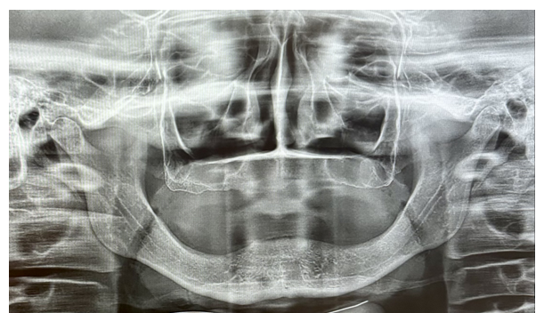
Figure 1: Preoperative panoramic view
FIGURE 2
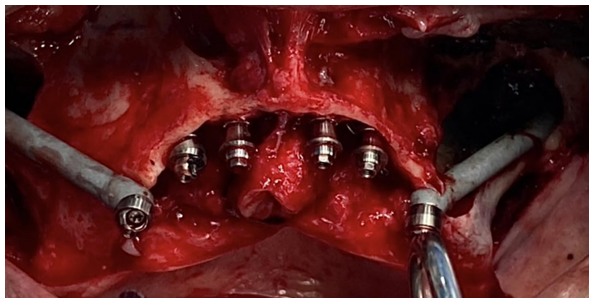
Figure 2: Zygomatic and endosseous dental implants
FIGURE 3
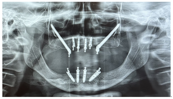
Figure 3: Postoperative panoramic view
FIGURE 4
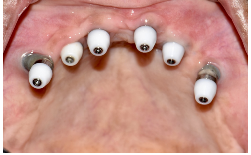
Figure 4: Healing abutments and implant positions
FIGURE 5
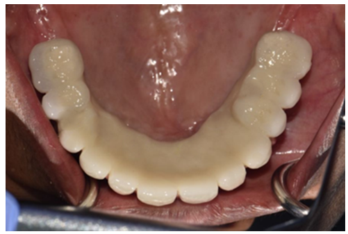
Figure 5: Completed upper prosthesis
FIGURE 6
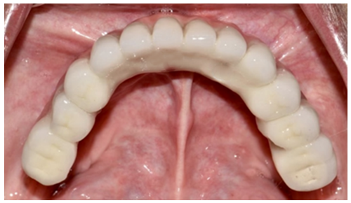
Figure 6: Completed lower prosthesis
FIGURE 7
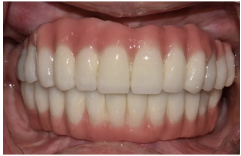
Figure 7: Completed upper-lower prosthesis
FIGURE 8
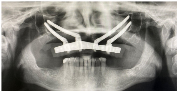
Figure 8: Panoramic view of a patient with Quad Zygoma application
Tables at a glance
Figures at a glance