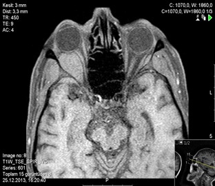
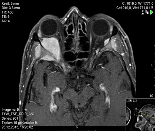
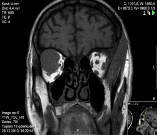
Figure 1: Bilateral orbital superiority in coronal T1WI is seen in the lateral aspect of the orbital and on the right side holding the large canal of the sphenoid bone and intraconal and extraconal left in the extracorporeal area gray matter and circumferential muscle tissue and isointense solid lesion.

Figure 2: In T1WI SPIR, there is no suppression due to lack of fat content in the lesions and isointense with gray matter

Figure 3: In T2WIretro bulbar fat is seen as isointense with the hypointense gray matter according to tissue

Figure 4: T2WIin the lesion does not contain oil in SPIR and is isointense with gray matter.

Figure 5: Diffusion restriction is observed in lesions

Figure 6: Contrast-enhanced T1WI SPIR shows a homogeneous contrast enhancement in the middle of the lesions.