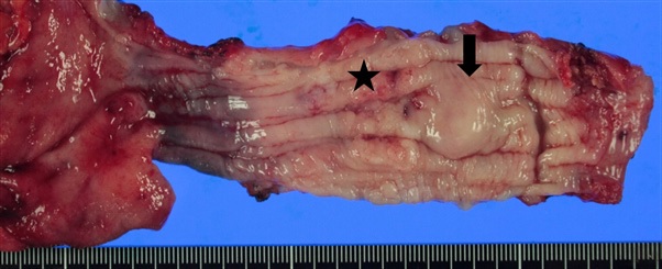
Figure.1a. An ulcerative lesion (asterisk) is found at 5cm away from the gastroesophageal junction. Submucosal lesion (arrow) is located 6.5cm away from the gastroesophageal junction.
Figure.1a. An ulcerative lesion (asterisk) is found at 5cm away from the gastroesophageal junction. Submucosal lesion (arrow) is located 6.5cm away from the gastroesophageal junction.
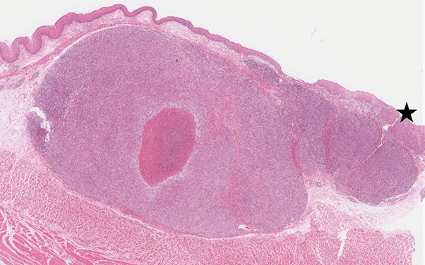
Figure.1b. The submucosal lesion is a relatively well-demarcated nodular pattern with central necrosis. Squamous carcinoma is also seen (asterisk). (H&E, X10)
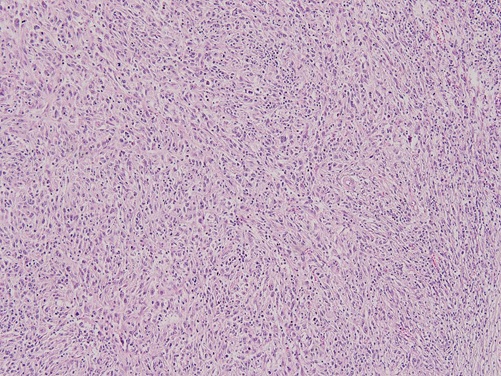
Figure.1c. The submucosal tumor is composed of interlacing fascicles of the short spindle to ovoid cells with vague storiform arrangement. The tumor cells show unequivocal nuclear atypism, prominent nucleoli, indistinct cell border, and a high mitotic figure representing more than 30 per 10 high power fields (HPFs). (H&E, X100)
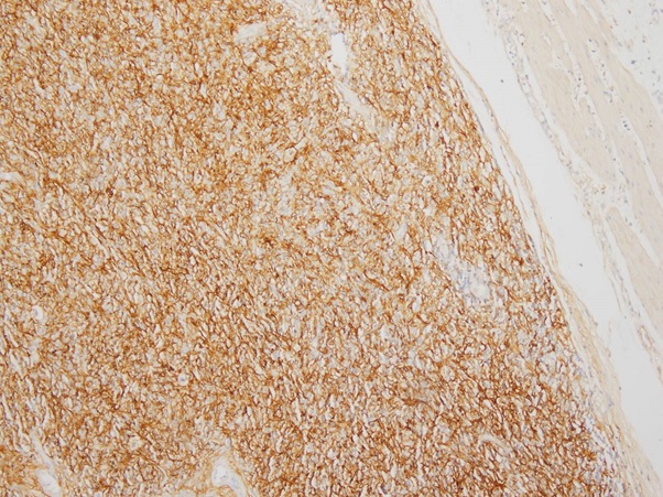
Figure.1d. The tumor cells show a strong immunoreactive for CD21. (X100)