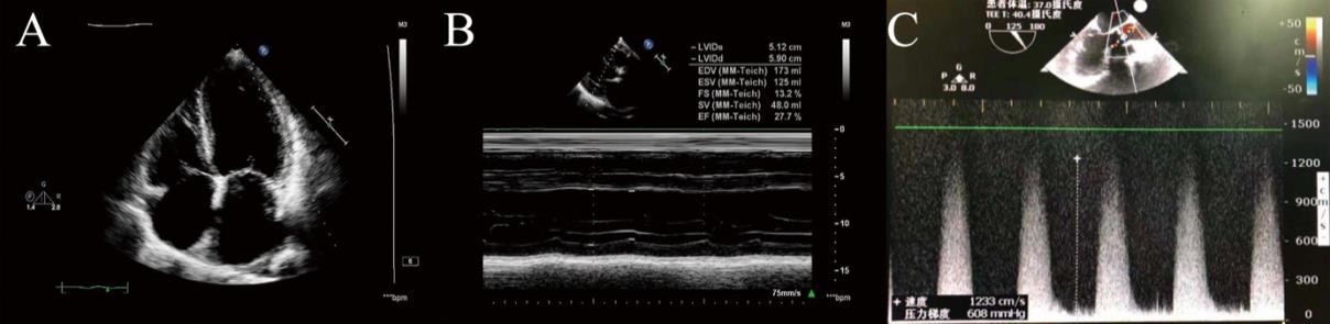
Figure 1: TTE and TEE findings before operation. (A-B) The preoperative TTE findings before operation; (C) The preoperative TEE findings before operation
Figure 1: TTE and TEE findings before operation. (A-B) The preoperative TTE findings before operation; (C) The preoperative TEE findings before operation
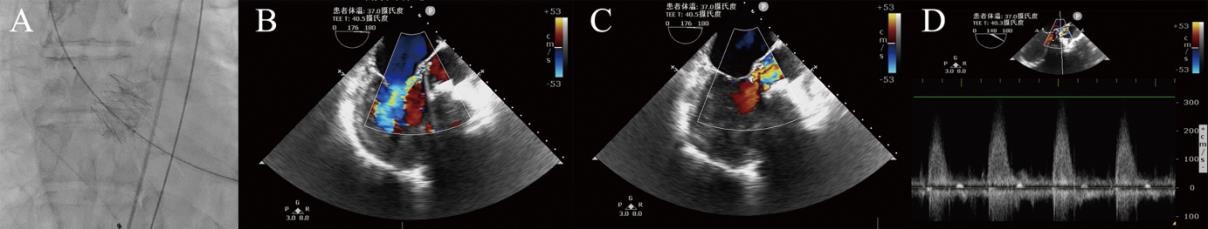
Figure 2: The fluoroscopic and TEE images after deployment of the prosthetic valve. (A) The fluoroscopic image after deployment of the prosthetic valve; (B-D) TEE images after deployment of the prosthetic valve
Figure 3: Thoracic HRCT scan showing lung involvement and the pleural sheath
Figures at a glance