Stajano, Fitz-Hugh, Curtis Syndrome. Review and Case Presentation
Received Date:January 09, 2023 Accepted Date: February 09, 2023 Published Date: February 13, 2023
doi: 10.17303/croa.2023.8.103
Citation: Fabián Rodríguez Escudero (2023) Stajano, Fitz-Hugh, Curtis Syndrome. Review and Case Presentation. Case Reports: Open Access 8: 1-8
Abstract
Stajano, Fitz-Hugh, Curtis Syndrome is an atypical clinical presentation of upper genital infections, characterized by few pelvic symptoms and perihepatitis determining right hypochondrium pain, tenderness, and "violin strings" hepatophrenic adhesions. This infrequent clinical presentation leads to frequent late or misdiagnoses, such as cholecystitis, appendicitis, urolithiasis, or hepatophrenic abscesses.
In this paper we carried out a historical review of knowledge of this particular clinical presentation.
Keywords: Ginecology; Stajano´s Syndrome; Phrenic reaction in gynecology; Fitz-Hugh; Curtis Syndrome; Chlamydia;Gonococcus
Background
Stajano, Fitz-Hugh, Curtis syndrome is an extrapelvic complication of pelvic inflammatory disease (PID), characterized by pain and tenderness in right hypochondrium secondary to perihepatitis, few pelvic symptoms, and presence of adhesions in “violin strings” forms between Glisson's capsule, diaphragm and anterior abdominal wall [1].
The clinical picture of a subacute or chronic gonococcal genital infection, which manifests as pain in right hypochondrium, with few pelvic symptoms, was first described by Dr. Carlos Stajano in 1920, publishing two articles in “Anales de la Facultad de Medicina del Uruguay” [2,3], the name given by Stajano to this clinical presentation was “phrenic reaction in gynecology” [3].
Few months later, Stajano published his findings in “La Semana Médica” of Buenos Aires magazine [4]. In 1921 JAMA published a summary and English translation of “La Semana Médica” article [5].
In 1922 Stajano published again in "Gynecologie et Obstetrique de Paris" [6] reaching wide dissemination in the European academic world, whose English summary and translation were published in 1922 by the "American Journal of Obstetrics & Gynecology" [7].
Ten years after the first description, in 1930, Dr. Arthur Hale Curtis published on the frequent coexistence of gonococcal salpingitis and “violin string” adhesions between anterior surface of liver and anterior abdominal wall [8].
In 1934, Dr. Thomas Fitz-Hugh Jr. published three cases of women who presented with pain in right hypochondrium due to adhesions between liver and abdominal wall, which he attributed to acute gonococcal peritonitis, linking his finding to the picture described by Curtis [9].
TIn 1978, Dr. Müller described the frequent occurrence in which Chlamydia trachomatis was isolated from peritoneal fluid in these clinical cases [10], in 1986 Lopez-Zeno linked this germ as the most frequently implicated agent [11], in 1970 Kimball and Knee published first case of male gonococcal perihepatitis [12], in 2003 Sharma et al. divulgated three cases as a result of genital tuberculosis [13] and cases secondary to Tuberculosis [14] and Mycoplasma have recently been published [15].
Clinical Case
A 27-year-old nulliparous patient with gynecological history of Pelvic Inflammatory Disease (PID) at 24 years of age treated with oral antibiotics. After this clinical picture, the patient frequently reports abnormal vaginal discharge that she self-medicates with vaginalsuppositories. During last year, she reported occasional pain in right hypochondrium interpreted as secondary to gallbladder disease, although imaging has not been able to diagnose gallstones or other anomalies that could explain this symptomatology.
Few months ago condition has worsened, and although it does not show pelvic symptoms, genital examination revealed tenderness on bimanual uterine palpation and cervical motion, and it has not responded satisfactorily to antibiotics treatment, analgesics and anti-inflammatories. It was decided to perform diagnostic and eventually therapeutic laparoscopy thinking on a Stajano, Fitz-Hugh, Curtis Syndrome.
During procedure, adhesions in a "violin string" form between liver and diaphragm, and fluid in Douglas cul-de-sac, and a genital sub-acute or chronic inflammatory process was observed. Figure 1
Adhesiolysis was performed, postoperative antibiotic treatment of PID is given. Patient had a satisfactory improvement and was discharge 48 hours after the procedure, follow up was uneventful.
Discussion
Stajano, Fitz Hugh, Curtis Syndrome is a rare form of chronic PID clinical presentation, giving with an inflammation of Glisson's capsule that finally causes perihepatic adhesions in "violin strings" form [1].
The patients often have pain in right hypochondrium with few pelvic symptoms. Occasionally pain may radiate to shoulder and is usually increased by Valsalva maneuvers; other times it is accompanied by fever, nausea and vomiting [1].
According to evolution, acute, sub-acute and chronic forms of presentation can be distinguished; most frequent were the last two [16].
During intra-abdominal exploration (laparoscopic or laparotomic), it demonstrates the “violin string” adhesions between liver and diaphragm, and less frequently with anterior abdominal wall [8]. Figure 1
The incidence is uncertain, although it is known that it frequently affects women of childbearing age who have suffered from PID. In the largest published study to date with 3,564 laparoscopies, this syndrome was diagnosed in 14.8 % of patients with tubal infertility; 6.7 % in ectopic pregnancies and in 1.4 % for other gynecological indications [17]. It is possible that this frequency is a little higher in adolescents since evidence of perihepatitis has been found in 27 % of patients with salpingitis [18], and the same it could happen in women with infertility [19].
The most frequent etiologic agent is Chlamydia trachomatis, followed by Neisseria gonorrhoeae [1,11].
Stajano has explained the reason why these genital infections adopt this curious clinical manifestation. A PID usually determines an endomyoparametritis, and if timely treatment is not given, the condition may evolve into a pyosalpinx, tube-ovarian abscess and pelviperitonitis. Sometimes, secondary to inadequate treatment, etiological agent characteristics or the patient's terrain, the infectious process evolves to a sub-acute or chronic stage. On these occasions, it may happen that the germs ascend from the pelvis, through the right parieto-colic gutter, to the hepatophrenic space [4,20,21]. This may be favored by the peritoneal fluid movements secondary to changes in intra-abdominal pressure caused by the inspiratory and expiratory efforts [21,22].
Once in the right hypochondrium, the inflammatory process produces a superficial hepatitis that almost exclusively affects Glisson's capsule, with production of loose adhesions, in a "violin strings" form, between it and diaphragm and anterior wall of hypochondrium [8,9]. Hematic and lymphatic have also been postulated as dissemination forms [23,24].
In a You et al. review it was reported that the most frequent symptom was pain in right hypochondrium (71 %), and later pain in hypogastrium (6.1 %), in right flank (4.9 %) and pleuritic pain (1.2 %) [25].
Stajano also explained why the pain usually occurs in right hypochondrium, and not in mid-abdomen. In upper abdomen, vegetative and sensory nerve endings are characterized by scarcity of underlying cellular tissue, so inflammatory process rapidly activates peritoneal pain receptors, while in peritoneal cavity middle level the nerve pathways cross muscle fibers, and ends in thicker connective and cellular tissue, which places them farther from peritoneal surface [20,21].
Although gynecological symptomatology is usually absent, it is rare that it does not manifest itself in some way during gynecological examination, both during speculoscopy (cervicitis, vaginal discharge), or during bimanual examination (pain on uterine palpation, cervix mobilization, adnexal, or pelviperitonitis) [1,3].
The “violin string” adhesions have usually been found as a laparoscopic finding, in a patient without suspicious symptoms. Tulandi and Falcone [26] published that at 4.7 % of laparoscopy performed on benign gynecological pathology, “violin string” adhesions can be found on the hepatic surface, which is consistent with that published by Ricci et al. [27]
This is why; it is always advisable to perform a cautious abdominal examination in women who undergo abdominal laparoscopy, regardless of the reason. If perihepatic adhesions are observed, adhesiolysis is recommended, which does not significantly increase the operating time or complications [28]. This practice usually improves symptoms and would avoid the complication derived from hemoperitoneum due to tearing of the hepatic capsule adhesions after abdominal trauma [29]. If lysis is not performed, it is advisable to record its presence and patient must have been warned, so as not to incur in future diagnostic errors [30,31]. Although more infrequent, there are also publications of cases of intestinal obstruction due to these adhesions [32,33].
If during a laparoscopy adhesiolysis is performed, great attention must be paid to hemostasis, since when abdominal pressure drops and when pneumoperitoneum is released, they may begin to bleed [34].
The blood test is the usual on PID, that is, leukocytosis with neutrophilia in half of the patients, and frequent elevation of C-reactive protein and erythrocyte sedimentation rate. Determination of antibodies against Chlamydia and Neisseria gonorrhoeae may be helpful although we rarely request it, the same happens with Chlamydia culture. Liver enzymes and bilirubin are usually normal, or slightly increased [1].
When a Nuclear Magnetic Resonance or a Computed Axial Tomography with contrast is performed, subcapsular fluid is usually observed, with hepatic capsule thickening in the arterial phase, secondary to the increase in blood flow caused by the inflammatory process. In 25 % of the patients it is possible to observe the adhesions in “violin strings” fashion, between hepatic surface and diaphragm and/or abdominal wall [35,36].
Given that clinical picture of Stajano, Fitz-Hugh, Curtis syndrome usually presents as pain in right hypochondrium, nausea and vomiting, and few pelvic symptoms, it is not uncommon to be attributed to hepatobiliary pathology, prompting misdiagnosis [1,30,31,37].
Antibiotic treatment [38] are the same as recommended in PID, and antibiotics used should be directed following the protocols established by each center, with special emphasis on treating the sexual partner(s).
Finally, regarding treatment, we believe it pertinent to distinguish 2 clinical situations: acute perihepatitis where surgical treatment is probably not necessary, thanks to antibiotic therapy, where second revision surgeries generally show regression of the lesions. And chronic or old perihepatitis, or when it coexists with infertility, where excision surgery may have a place.
Conclusion
Stajano, Fitz-Hugh, Curtis syndrome is a rare clinical presentation of upper gynecological infections, characterized by right hypochondrium pain, few pelvic symptoms, and adhesions in a "violin string" form between liver and diaphragm or anterior abdominal wall.
Because clinical characteristics, diagnosis is usually delayed or is erroneously made, its usually being assigned to hepatobiliary, appendicular or urological pathology.
The blood test is nonspecific, with a slight leukocytosis with neutrophilia, increased CRP and SED rate. Liver enzymes are usually normal or slightly increased.
It is also not uncommon for “violin string” adhesions to be a surgical finding in an asymptomatic patient.
Treatment consists of laparoscopic adhesiolysis, and antibiotic therapy typical of PID.
- Gómez A, Parras F, Quesada MN, Troncoso AC, Sánchez JJ et al. (2018) Síndrome de Fitz-Hugh-Curtis como causa de dolor agudo en hipocondrio derecho en paciente en edad fértil. Prog Obstet Ginecol 61: 594-8.
- Pouey E, Stajano C (1920) Las reacciones del peritoneo supracéliaco en ginecología, Anales Facultad Medicina 5: 202.
- Stajano C (1920) La reacción frénica en ginecología e interpretación fisiológica. Anales Facultad Medicina 5: 722.
- Stajano C (1920) La reacción frénica en ginecología. Semana Médica (Buenos Aires) 27: 243-8.
- “Semana Médica, Buenos Aires (1921) Phrenic reaction in gynecology. C Stajano.” Current Medical Literature. JAMA 76: 690.
- Stajano C (1922) Reaction phrénique et infection genitale. Gynecologie et Obstétrique, París 1: 42-8.
- Adair F (1923) “Diaphragmatic reaction in Genital infection: Stajano C: Gynecologie te Obstetrique, 1922, VI, 44”. Amer Jour Obstet & Gynecol 6: 508.
- Curtis AH (1930) A cause of adhesions in the right upper quadrant. JAMA 94: 1221-2.
- Fitz-Hugh T Jr (1934) Acute gonococcic peritonitis of the right upper quadrant in women. JAMA 102: 2094-6.
- Müller J, Wang S, Munzinger J et al. (1978) Chlamydia trachomatis as a possible cause of peritonitis and periheptitis in young women. Br Med J 1: 1002-4.
- Lopez-Zeno JA, Keith LG, Berger GS (1985) The Fitz-Hugh-Curtis syndrome revisited. Changing perspectives after half a century. J Reprod Med 30: 567-82.
- Kimball MW, Knee S (1970) Gonococcal perihepatitis in a male. The Fitz-Hugh-Curtis síndrome. The New England Journal of Medicine 282: 1082-4.
- Sharma JB, Malhotra M, Arora R (2003) Fitz-Hugh-Curtis syndrome as a result of genital tuberculosis: a report of three cases. Acta Obstet Gynecol Scand 82: 295-7.
- Coremans L, de Clerck F (2018) Fitz-Hugh-Curtis syndrome associated with tuberculous salpingitis and peritonitis: a case presentation and review of literature. BMC Gastroenterol 18: 42.
- Flores-Sánchez I, GutierrezSalinas J, Calderón-Tapia J, Cervantes-Chávez JF (2011) Síndrome de Fitz-Hugh-Curtis asociado con Mycoplasma Hominis: caso clínico. Ginecol Obstet Mex 10: 637-41.
- Demelo P, González A, Tejerina F, Bernaldo JC (2013) Síndrome de Fitz-Hugh-Curtisen fase subaguda/crónica. Rev Clin Esp 213: e49.
- Hong DGMH, Choi MH, Chong GO, Yi JH et al. (2010) Fitz-HughCurtis Syndrome: Single centre experiences. J Obstet Gynaecol 30: 277-80.
- Litt IF, Cohen MI (1978) Perihepatitis associated with salpingitis in adolescents. JAMA 240: 1253-4.
- Godinjak Z, Idrizbegovic E (2008) Should diagnostic hysteroscopy be a routine procedure during diagnostic laparoscopy in infertile women? Bosn J Basic Med Sci 8: 44-7
- Stajano C (1930) Las leyes del dolor visceral. Soca año II N°17.
- Stajano C (1931) Fisiopatología clínica del dolor en ginecología. Soca año II 18: 11
- Hunter RH, Cicinelli E, Einer-Jensen N (2007) Peritoneal fluid as an unrecognized vector between female reproductive tissues. Acta Obstet Gynecol Scand 86: 260-5.
- Hamdan M, Johanet H, Benhamou G (1992) The Fitz-Hugh-Curtis syndrome in a man revealed by ectopic appendicitis. Eur J Med 1: 314-5.
- Davidson AC, Hawkins DA (1982) Pleuritic pain: Fitz-Hugh-Curtis syndrome in a man. Br Med J (Clin Res Ed) 284: 808.
- You JS, Kim MJ, Chung HS, Chung YE, Park I et al. (2012) Clinical features of fitz-hugh-curtis syndrome in the emergency department. Yonsei Med J 53: 753-8.
- Tulandi T, Falcone T (1998) Incidental liver abnormalities at laparoscopy for benign gynecologic conditions. J Am Assoc Gynecol Laparosc 5: 403-6.
- Ricci P, Lema R, Solá V et al. (2008) Fitz-Hugh-Curtis syndrome: Three cases of incidental diagnosis during laparoscopy. J Obstet Gynaecol 28: 352-4.
- Palade R, Vasile D, Grigoriu M, Voiculescu D (2002) The Fitz-Hughes-Curtis síndrome in laparoscopic surgery. Chirurgia (Bucuru) 97: 557-61.
- Foster HM (1998) Haemoperitoneum: An unusual complicaction of Fitz-Hugh Curtis syndrome. Aust N Z J Surg 58: 342-3.
- Woo SY, Kim Ji, Cheung DY, Cho SH, Park SH et al. (2008) Clinical outcome of Fitz-Hugh-Curtis syndrome mimicking acute biliary disease. World J Gastroenterol 14: 6975-80.
- Piscaglia F, Vidili G, Ugolini G, Ramini R, Montroni I et al. (2005) Fitz-Hugh-Curtis mimicking acute cholecystitis: value of new ultrasound findings in the differential diagnosis. Ultraschall Med 26: 227-9.
- Burton E, McKeating J, Stahlfeld K (2008) Laparoscopic management of a small bowel obstruction of unknown cause. JSLS 12: 299-302.
- Abul-Khoudoud OR, Khabbaz AY, Butcher CH, Farha MJ (2001) Mechanical partial bowel obstruction in a patient with Fitz-Hugh-Curtis syndrome. J Laparoendosc Adv Surg Tech A 11: 111-4
- Rogers RG, Monahan EG (1996) Postoperative hemorrhage due to avulsion of perihepatic adhesions after pneumoperitoneum. J Am Assoc Gynecol Laparosic 3: 631-3.
- Cho HJ, Kim HK, Suh JH, Lee GJ, Shim JC et al. (2008) Fitz-HughCurtis syndrome: CT findings of three cases. Emerg Radiol 15: 43-6.
- Wang CL, Guo XJ, Yuan ZD, Shi Q, Hu XH et al. (2009) Radiologic diagnosis of Fitz-Hugh-Curtis syndrome. Chin Med J (Engl)122: 741-4.
- Peter NG, Clark LR, Jaeger JR (2004) Fitz-Hugh-Curtis syndrome: a diagnosis to consider in women with right upper quadrant pain. Cleve Clin Med 71: 233-9.
- Ross J, Guaschino S, Cusini M, Jensen J (2018) Guía europea para el manejo de la Enfermedad Inflamatoria Pelviana. Int Jour STD&AIDS 29: 108-14.
FIGURE 1
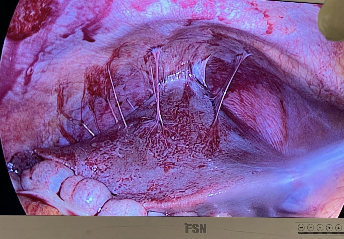
Figure 1: Hepatophrenic adhesions in the "violin strings" form, in a patient with Stajano, Fitz-Hugh, Curtis Syndrome. Photo courtesy of Dr. Gonzalo Sotero
Figures at a glance