
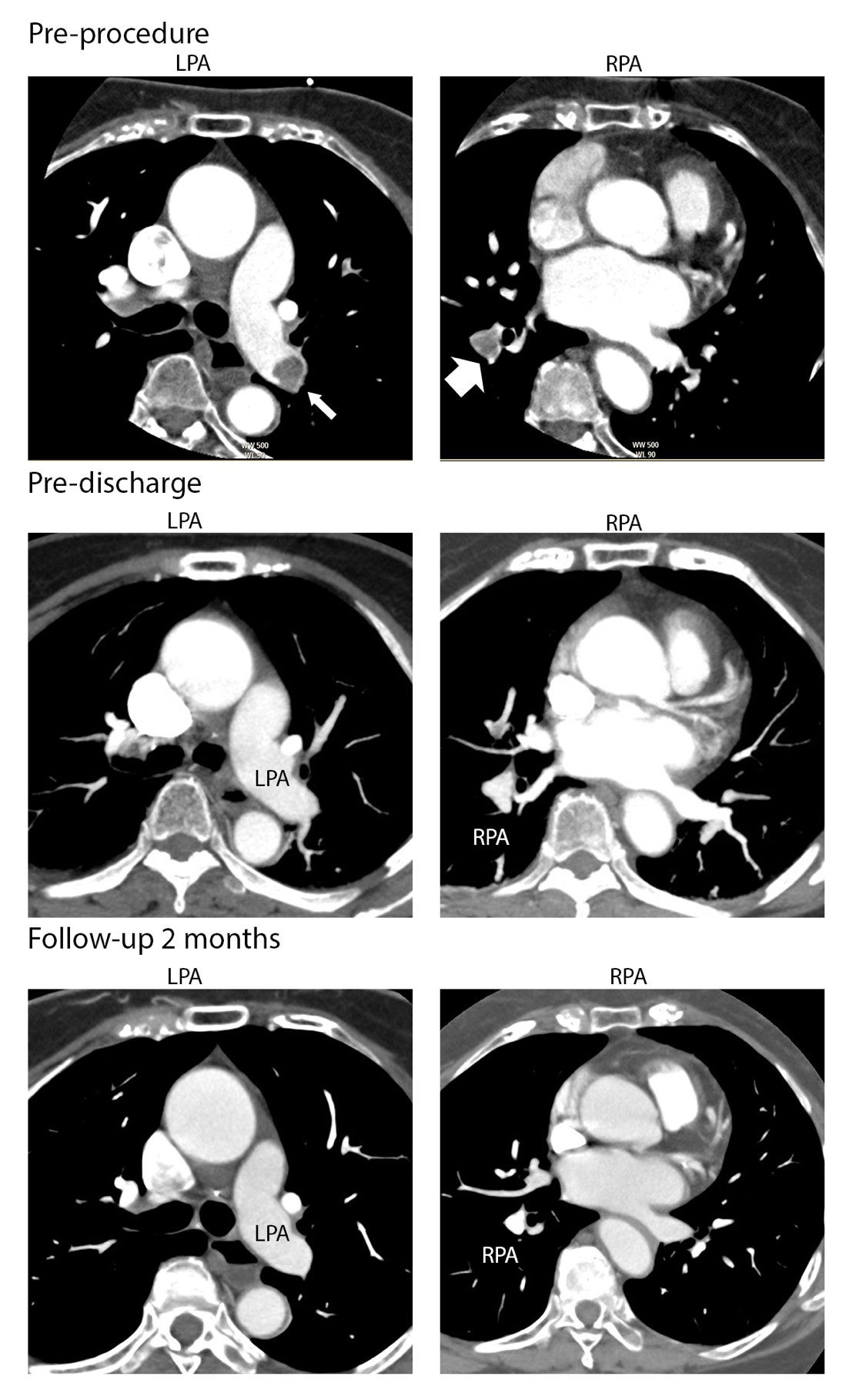
Figure 1: [Pre-procedure] CTPA showing a large thrombus covering the right PA (big arrow) and left inferior segment of the PA (small arrow). [Pre-discharge] CTPA showing residual thrombus in the inferior segment of left PA. [Follow-up 1 month] CTPA showed increasing thrombus size at the inferior segment of left PA and minimal thrombus at the inferior segment of right PA. [Follow-up 3 months] CTPA showing complete thrombus resolution at the right PA and left inferior segment of PA
Figures at a glance