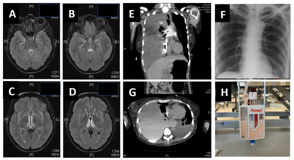
Figure 1:A-D: FLAIR-weighted MRI showing typical, symmetric signal enhancement for Wernicke encephalopathy from mamilary bodies up to both thalami. E,G: CT thorax showing bilateral pleural effusion with massive accentuation on the right side, mild pericardial effusion and mediastinal shift. F: Thoracical X-ray showing correct placement of the subclavian line from left side. H: Fluid collected by pleural drainage.
Figures at a glance