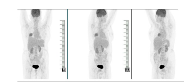
Figure 1: PET-CT done in 2019 at the time of presentation
Figure 1: PET-CT done in 2019 at the time of presentation
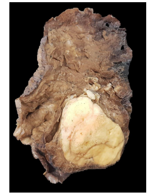
Figure 2: Gross specimen post lobectomy
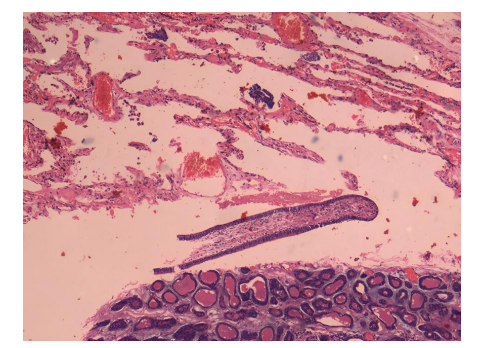
Figure 3: Low power view showing lung parenchyma with the attached tumor
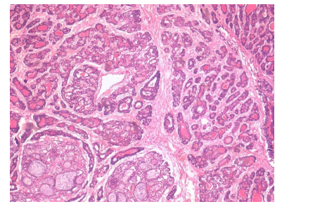
Figure 4: 200 x Tumor cells showing cribriform pattern composed of inner ductal and outer myoepithelial cells
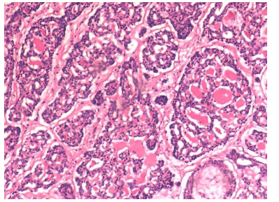
Figure 5: 400 x showing higher power view of tumor cells confirming the adenoid cystic tumor
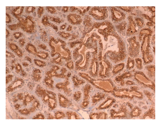
Figure 6: IHC for CD117 confirms the diagnosis of adenoid cystic carcinoma
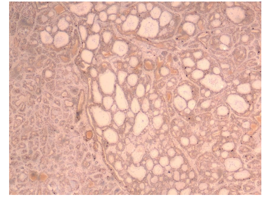
Figure 7: IHC for Ki67 proliferation index less than 2%
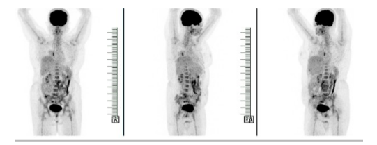
Figure 8: PET-CT done in 2020 during annual follow-up: Focal non FDG avid air space opacity noted in the right perihilar region,FDG avid foci are seen in the right lower lobe lobectomy stump
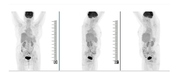
Figure 9: PET-CT done in 2021: No recurrence/metastasis
Figures at a glance