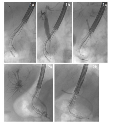
Figure 1: 1a-c representing the case of a patient with a distal cholangiocellular carcinoma depicting wire cannulation (a) positive cholangiogram (b) cholangioscopy (c) 2a-b representing a case of a patient with Klatskin III cancer depicting the positive cholangigramm (2a) and cholangioscopy (2b)
Tables at a glance
Figures at a glance