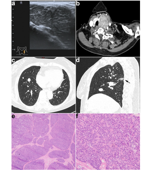
Figure 1A: Ultrasound showed that the left thyroid had a large hypoechoic solid nodule with a clear margin. (B) Enhanced-CT in the axial plane showed an irregular heterogeneous enhanced mass within the left thyroid pushing the trachea to the right. (C) Non-enhanced CT in the axial plane showed multiple solid nodules in the inferior lobe of the right lung. (D) Non-enhanced CT in the sagittal plane showed a mixed ground-glass opacity in the dorsal part of the inferior lobe of the right lung (arrow) and a solid nodule. (E) In the frozen section, the tumor showed a multinodular growth pattern characteristic of the "jigsaw puzzle" pattern. (F) At a higher magnification of (E), tumor cells formed a pseudo-glandular pattern
Figures at a glance