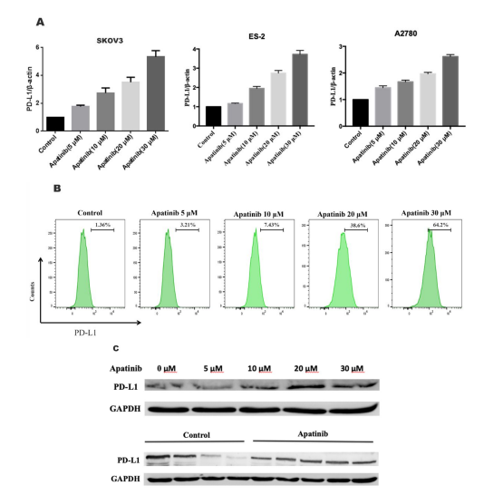
Figure 1: Apatinib increased PD-L1 expression in ovarian cancer cells. a SKOV-3, ES-2, and A2780 ovarian cancer cells were treated with different concentrations of apatinib for 24 h, and PD-L1 mRNA expression was detected via RT‒PCR. b SKOV-3 cells were treated with different concentrations of apatinib for 24 h, and PD-L1 protein expression was detected via flow cytometry and Western blotting. c SKOV3 (5 × 10⁶ ) cells were inoculated into the right flank of BALB/c mice. Mice were administered apatinib (30 mg/kg, every day). After 15 days, the mice were sacrificed, and their solid tumours were isolated. The expression of PD-L1 in the tumour sections was analysed via western blotting. The results are expressed as the mean ± SEM from three independent experiments. *P < 0.05, **P < 0.01 vs. control.
Figures at a glance