A large Posterior Mediastinal Cavernous Lymphangioma with Benign Osteolysis Foci: A Case Report
Received Date: May 24, 2024 Accepted Date: June 24, 2024 Published Date: June 27, 2024
doi: 10.17303/jcrto.2024.12.302
Citation: Othman Hamdan, Balqees Al-Shohef, Nagham Al-Shohef (2024) Department of Hematology, Children's University Hospital, Damascus University, Damascus, Syria. J Cancer Res Therap Oncol 12: 1-9
Abstract
Background: Lymphangioma is a rare benign malformation of lymphatic system that is often diagnosed in the first few years of life. Occurs due to blockage of the lymphatic system during fetal development. They commonly occur in the neck, axilla, and extremities [1,2].
Keywords: Mediastinal; Cavernous; Lymphangioma; Posterior
Introduction
We report a case of a Syrian child with a large cavernous lymphangioma in posterior mediastinum presenting with: dyspnea, recurrent pneumonia, fever.
Case Presentation
A 5-year-old Syrian boy presented with recurrent pneumonia, fever, dyspnea. No weight loss or night diaphoresis. On examination he was afebrile, and had supcostal, intercostal, and suprasternal
recessions, with respiratory rate of 30 breaths per minute. He had aloud respiratory grunt. The chest expansion was reduced which was dull on percussion. Asculation revealed a marked reduction of air entry over the mid and upper zones. Chest-X ray showed a well demarcated opacity involving the upper and mid zones. Ultrasound scan of the chest revealed fluid-filled right costophrenic angle, suggesting a pleural effusion.
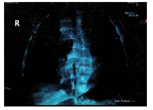
A contrast-enhanced Computer Tomography Scan of the chest showed: a large mass filling the posterior mediastinum in it's upper and middle parts, aligned with the visceral side of the right upper lobe.
MRI showed: a mass in the posterior mediastinum surrounding the tracheal branch and reaches the diaphragm, surrounding the Azygos vein, superior vena cava, aorta, major vessels and esophagus, measuring (11*5*7) cm. and foci osteolysis in the 9,11,12 thoracic vertebrae.
His full blood count revealed: white cell count of 15.000/mm³, neutrophils 10.500/mm³, lymphocytes 4500/mm³, eosinophils 300/mm³, hemoglobin of 12.8 g/dl and platelets of 333.000/mm³. His C- reactive protein was 6mg/l. His serum electolytes, renal and liver function were normal. Alpha- fetoprotein, and neuron-specific enolase (NSE) were also normal.
Peripheral blood smear: normal.
Bone marrow aspiration: normal.
Bone marrow biobsy: benign bone marrow.
Bone scanning: hypodense foci on the left and right thight, from which biopsies were taken and the bone tissue were normal, with no malignancy.
Bone scintigraphy: foci of enhacement of the material (radioactive Technetium) in the distal third of the thight, with increased fixation of the radioactive material in the posterior end of the eight vertebra, no metasis.
Discussion
Lymphangiomas are uncommon benign malformations first described in 1913 by Gaudier and Gorse. Occur due to blockage of the lymphatic system during fetal development [3,4].
Conginital form typically occurs before the age of 5years old [5].
Due to dissconection of lymphatic channels to the main lymphatic drainage duct.
Though acquired lymphangioma occur as a result of any interruption of previously normal lymphatic drainage such as: trauma, malignancy, surgery, and radiation therapy and presenting in adulthood [6].
Our patient has the congenital form and he initially underwent drainage of the pleural effusion with antibiotic treatment, and biopsies of bone foci and lungs were accomplished:
mained symptoms free at 6-weeks to 3-month follow-up visits. He was on conservative treatment due to the location of the mass which surrounding the vessels, in addition to the difficulty and high risk of the surgical operation.
With an annual periodic review, for blood tests and a chest CT scan that showed a decrease in the size of the lymphangioma.
Lung biopsy: pulmonary tissue.
Mass biopsy: no granuloma, no malignancy or TB, cavernous lymphangioma.
And the diagnosis was confirmed.
The child showed a complete recovery and he remained symptoms free at 6-weeks to 3-month follow-up visits. He was on conservative treatment due to the location of the mass which surrounding the vessels, in addition to the difficulty and high risk of the surgical operation
With an annual periodic review, for blood tests and a chest CT scan that showed a decrease in the size of the lymphangioma
Conclusion
We report a case with a rare occurrence of a large cavernous lymphangioma originating from posterior mediastinum. It highlights the importance of considering rare possibilities and performing imaging in uncertainly diagnosed situations that can be life-saving.
- Rathan JJ, Vardhan BG, Muthu MS, Venkatachalapathy, Saraswathy K, Sivakumar N (2005) Oral lymphangioma: A case report. J Indian Soc Pedod Prev Dent. 23: 185-9.
- Mentzel HJ, Schramm D, Vogt S, Reuter A, Mentzel T, Kaiser WA (1998) Intra-abdominal lymphangioma in a newborn. J Clin Ultrasound, 26: 320-2.
- Ersoy AO, Oztas E, Saridogan E, Ozler S, Danisman N (2016) An Unusual Origin of Fetal Lymphangioma Filling Right Axilla. J Clin Diagn Res. 10: QD09-11.
- Kosir MA, Sonnino RE, Gauderer MW (1991) Pediatric abdominal lymphangiomas: a plea for early recognition. J Pediatr Surg, 26: 1309-13.
- Sehgal VN, Sharma S, Chatterjee K, Khurana A, Malhotra S (2018) Unilateral, Blaschkoid, Large Lymphangioma Circumscriptum: Micro- and Macrocystic Manifestations. Skinmed. 16: 411-3.
- Zadvinskis DP, Benson MT, Kerr HH, Mancuso AA, Cacciarelli AA, et al. (1992) Congenital malformations of the cervicothoracic lymphatic system: embryology and pathogenesis. Radiographics, 12: 1175-89.
FIGURE 1
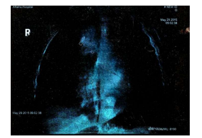
FIGURE 2
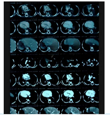
FIGURE 3
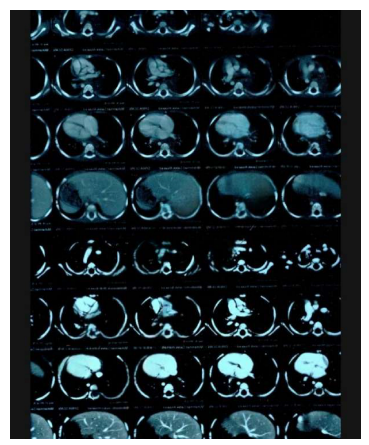
FIGURE 4
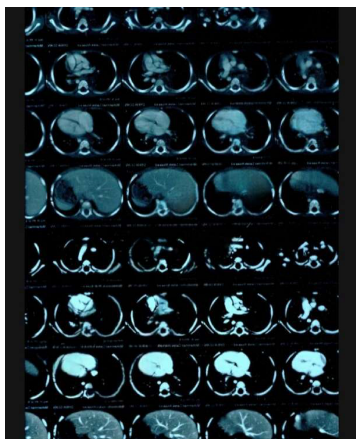
FIGURE 5
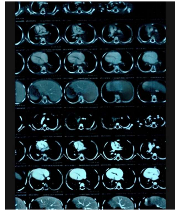
FIGURE 6
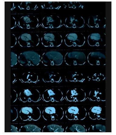
FIGURE 7
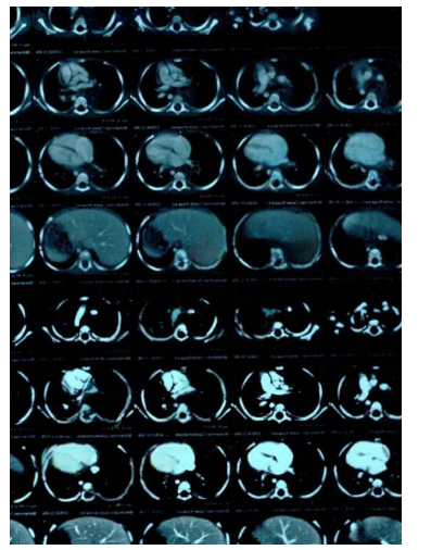
FIGURE 8
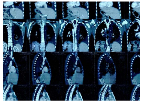
FIGURE 9
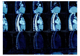
FIGURE 10
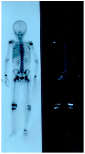
FIGURE 11
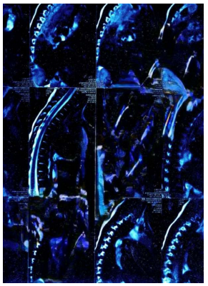
FIGURE 12
Figures at a glance