Table 1: Clinical Characteristics and Treatment Regime of Patients.
CT: chemotherapy; RT: Radiation therapy; ICT: Induction chemotherapy; CCT: Concurrent chemotherapy; ACT: Adjuvant chemotherapy; PCT: Palliative chemotherapy; IMRT: Intensity-modulated radiation therapy; WBRT: Whole-brain radiation therapy
Table 2: Literature Review.
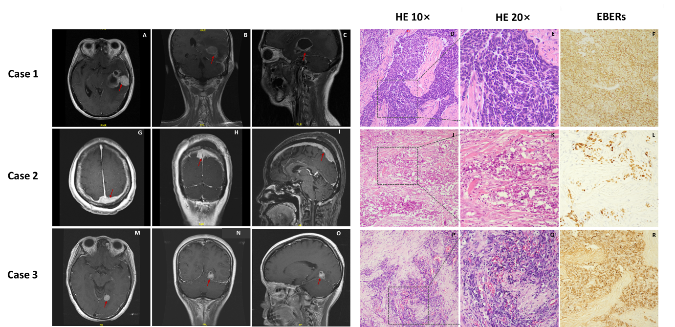
Figure1: Magnetic resonance and pathological images of patients.
Tables at a glance
Figures at a glance