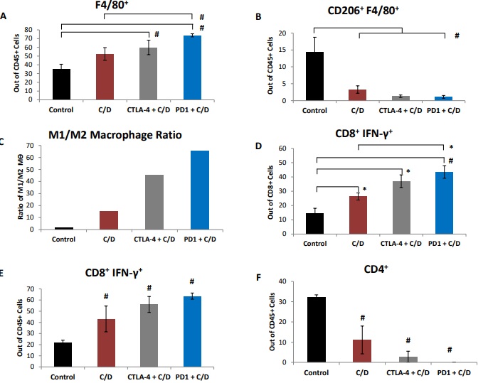
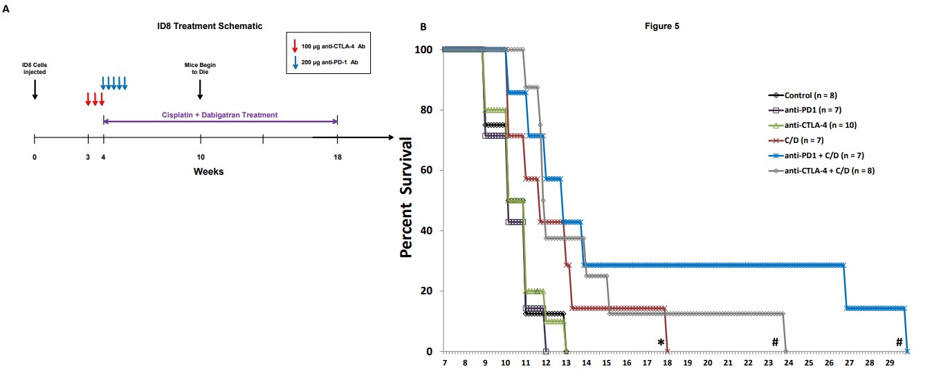
Figure 1 Co-treatment with cisplatin and dabigatran etexilate reduces tumor burden early during tumor progression. Four weeks after mice were i.p. injected with 2.0 x 10⁶ ID8-luc cells, all treatments were initiated. Anti-PD-1 antibodies (200 μg every two days, five doses total) were i.p. injected. Mice were injected i.p. with cisplatin (1 mg/kg) once weekly. Dabigatran etexilate was administered by oral gavage twice daily (80 mg/kg) Monday through Friday, and mice were placed on chow supplemented with dabigatran etexilate (10 mg/g chow) over the weekends. (A) Final tumor loads of mice assessed by bioluminescence imaging of the opened peritoneal cavity six weeks after tumor injection (2 weeks of treatment with cisplatin and dabigatran). (B) Cells (2 x10⁶) isolated from peritoneal lavages were treated for 4 h at 37ºC with ionomycin (500 ng/ml) and PMA (50 ng/ml) to stimulate activated T cells to produce IFN-γ in the presence of Brefeldin A to block cytokine secretion. Ascites cells were stained with anti- CD8α and anti-CD45 antibodies in the presence of Brefeldin A. Cells were then fixed, permeabilized, and intracellularly stained with an anti-IFN-γ antibody and analyzed by flow cytometry. n = 5 mice per group ± SEM; * = p< 0.05 and # = p<0.01 compared to control vehicle-treated tumor-bearing mice.

Figure 2 Inhibitory effects of cisplatin and dabigatran etexilate co-treatment plus either anti-CTLA-4 or anti-PD-1 therapy on ID8 tumor growth and malignant ascites accumulation. Mice were treated as described in Figure 1, apart from anti- CTLA-4 treated mice. Mice in the CTLA-4 groups received three doses of anti-CTLA-4 antibodies starting at week three after ID8 cell injection every third day for three doses. All mice were sacrificed after six weeks of treatment (10 weeks after tumor cell injection).(A) Representative quantification of ID8-luc tumor burden by bioluminescence imaging in living mice. (B) Ascites volume was determined upon sacrifice. (C) The final tumor loads were assessed by bioluminescence imaging of the opened peritoneal cavity. Insert is an enlarged figure of bioluminescence for mice treated with C/D ± immune checkpoint inhibitors. (D) Representative quantification of ID8-luc tumor burden by bioluminescence imaging in living mice. Mice were sacrificed at 10 weeks post tumor injection. n = 7-10 mice per group ± SEM; * = p< 0.05 and # = p< 0.01 compared to control vehicle-treated tumor-bearing mice.

Figure 3 Cisplatin and dabigatran etexilate co-treatment plus either anti-CTLA-4 or anti-PD-1 therapy increases cytotoxic T-cell IFN-γ production and decreases M2 macrophage levels in the ascites ID8-luc tumor-bearing mice. Upon sacrifice, ascites was removed from ID8 tumor-bearing mice and spun at 300 g for 10 minutes to isolate the cellular component. (A-B) CD45+ ascites cells were stained and analyzed by flow cytometry for the percentage of the indicated leukocytes. (C) The ratio of pro-inflammatory M1 macrophages to pro-tumorigenic M2 F4/80+CD206+ macrophages where M1 macrophages are [(total F4/80+ macrophages) – (F4/80+CD206+ M2 macrophages)]. (D) Ascites cells (2 x 10⁶) were treated for 4 h at 37ºC with ionomycin (500 ng/ml) and PMA (50 ng/ml) to stimulate activated T cells to produce IFN-γ in the presence of Brefeldin A to block cytokine secretion. Ascites cells were stained with anti- CD8α and anti-CD45 antibodies in the presence of Brefeldin A. Cells were then fixed, permeabilized, and intracellularly stained with an anti-IFN-γ antibody and analyzed by flow cytometry. (E-F) Upon sacrifice, ID8 tumors on the peritoneal wall of tumor-bearing mice were excised, digested and processed into a single cell suspension for analysis by flow cytometry. CD45+ cells were analyzed for the percentage of the indicated tumor-infiltrating leukocytes. n = 7-10 mice per group ± SEM; * = p< 0.05 and # = p< 0.01 compared to control vehicle-treated tumor-bearing mice or indicated groups.

Figure 4 Cisplatin and dabigatran etexilate co-treatment plus either anti-CTLA-4 or anti-PD-1 therapy reduce levels of pro-tumorigenic cytokines in the ascites. Upon sacrifice, ascites were removed and spun at 300 g for 10 minutes to isolate the cell-free component of the ascites which was assayed for levels of TNF-α, MCP-1, IL-10, IL- 6, and TGF-β by Cytokine Bead Array. n = 7-10 mice per group ± SEM; * = p< 0.05 and # = p< 0.01 compared to control vehicle-treated tumor-bearing mice or indicated groups.

Figure 5Co-treatment with cisplatin and dabigatran etexilate plus either anti- CTLA-4 or anti-PD-1 therapy confers a survival benefit in mice with ID8 ovarian tumors. (5A) Overall schematic of ID8 ovarian tumor survival experiments. Three weeks after mice were i.p. injected with 2.0 x 10⁶ ID8-luc cells, administration of anti- CTLA-4 antibodies (100 μg every three days, three doses total) was initiated. At week 4, all other treatments were initiated. Anti-PD-1 antibodies (200 μg every two days, five doses total) were i.p. injected. Mice were injected i.p. with cisplatin (1 mg/kg) once weekly. Dabigatran etexilate was administered by oral gavage twice daily (80 mg/kg) Monday through Friday, and mice were placed on chow supplemented with dabigatran etexilate (10 mg/g chow) over the weekends. Treatment with cisplatin and dabigatran etexilate (C/D) was terminated at week 18. Figure 5B: Kaplan-Meier survival curves of ID8 tumor-bearing mice treated with vehicle, anti-CTLA-4 alone, anti-PD-1 alone, C/D with or without anti-CTLA-4, or C/D with or without anti-PD-1. n = 7-10 mice per group; * = p<0.05 and # = p<0.01 compared to control vehicle-treated tumor-bearing mice.

Supplementary Figure 1.Upon sacrifice, ascites was removed from ID8 tumor-bearing mice, and the cellular component was isolated. (A-B) CD45+ ascites cells were gated and analyzed by flow cytometry for the percentage of the indicated CD8+ or CD4+ T-cells. n = 7-10 mice per group ± SEM; * = p<0.05 and # = p<0.01 compared to control vehicle-treated tumor-bearing mice or indicated treatment groups.