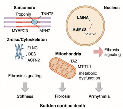
Figure 1: Molecular pathways and cellular structures are affected in RCM
This schematic illustrates the key molecular components and cellular structures implicated in the pathogenesis of restrictive cardiomyopathy. The sarcomere is shown with contractile proteins including MYH7, MYBPC3, and troponins (TNNI3, TNNT2), reflecting impaired myocardial contraction. The Z-disc/cytoskeleton is represented by FLNC, DES, and ACTN2, proteins critical for structural integrity and mechanotransduction. The nucleus features LMNA and RBM20, linked to nuclear envelope stability and transcriptional regulation. Mitochondrial dysfunction is illustrated by TAZ and MT-TL1, associated with impaired energy metabolism. The fibrotic response is highlighted through the TGF-β signaling pathway, a central mediator of myocardial fibrosis. Arrows depict downstream pathological consequences including increased myocardial stiffness, fibrotic remodeling, arrhythmias, and the risk of sudden cardiac death.
Tables at a glance
Figures at a glance