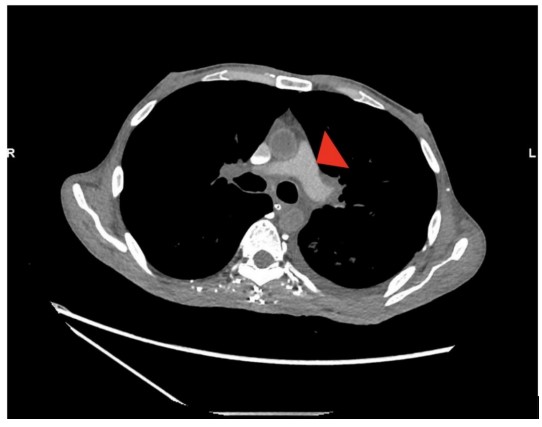
Figure 1: Poor opacification of the pulmonary trunk following the test bolus is annotated by an arrow head.
Figure 1: Poor opacification of the pulmonary trunk following the test bolus is annotated by an arrow head.
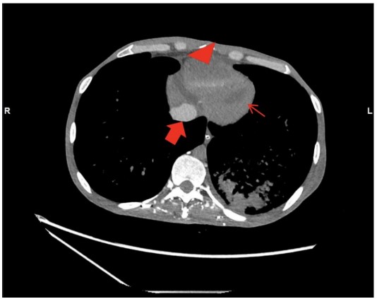
Figure 2: Dependent layering of contrast within the right ventricle is annotated by a red arrow head. Contrast pooling within inferior vena cava is annotated by a thick red arrow. Absent of contrast in the left ventricle and aorta is annotated by thin red arrows.
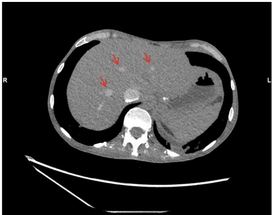
Figure 3: Pooling of contrast material in the hepatic veins annotated by thin red arrows.
Figures at a glance