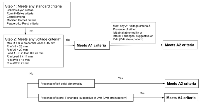
Figure 1: Stepwise algorithm for ECG-based LVH detection incorporating standard and additional (A1–A4) criteria
* A1–A4 represent additional criteria applied only when standard criteria are not met.
Figure 1: Stepwise algorithm for ECG-based LVH detection incorporating standard and additional (A1–A4) criteria
* A1–A4 represent additional criteria applied only when standard criteria are not met.
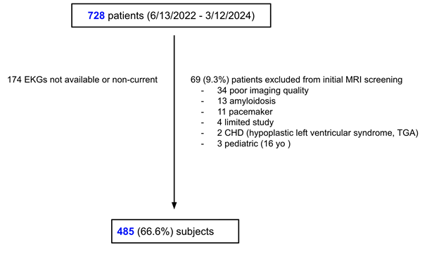
Figure 2: Flowchart of patient enrollment
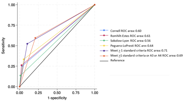
Figure 3: ROC curve analysis of ECG-LVH criteria for detecting elevated LV mass index. A3: represents left atrial enlargement (LAE), A4: represents lateral T-wave changes.
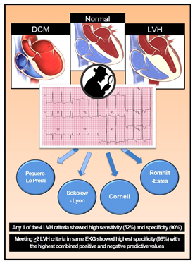
Figure 4: Central illustration. DCM: Dilated Cardiomyopathy, LVH: Left Ventricular Hypertrophy.
Tables at a glance
Figures at a glance