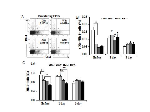
Figure 1: Circulating EPCs before and after arterial injury
A, Representative histograms of flow cytometry analysis of circulating c-Kit+/ Flk-1+ cells, which were significantly increased in the blood of Dki mice. B and C, Kinetic alterations of circulating c-Kit+/Flk-1+ cells and Flk-1+ cells before and after injury at the indicated time points, determined by flow cytometry. *P< 0.05; **P< 0.01, vs. indicated groups; #P< 0.05 vs. EPCs level before injury (n=4-7/group).

Figure 2: EPC mobilization pathway in bone marrow: MMP-9 and sKitL levels
A, Detection of bone marrow MMP-9 activity without arterial injury by gelatin zymography. B, Bone marrow plasma soluble Kit ligand (sKitL) levels before and after injury were determined with ELISA and normalized with total protein contents (100 μg). *P< 0.05; **P< 0.01; #P< 0.05 vs. indicated groups (n=4-7 mice/group).

Figure 3: Effects of eNOS on proliferation and apoptosis of circulating EPCs
Proliferation (A) and apoptosis (B) of circulating Flk-1+ cells detected by cell cycle analysis and Annexin V/PI double staining. **P< 0.01, vs. WT group (n=5-8/group). C, Representative histograms of flow cytometry analysis of activation of caspase-3 in circulating Flk-1+ cells. and quantification of active caspase-3+ cells in total Flk-1+ cells. *P< 0.05 vs. WT group (n=5-6 mice/ group).

Figure 4: Incorporation of bone marrow-derived circulating EPCs into regenerated endothelium
A, Experimental protocol. B, FACS analysis indicates that bone marrow in four strains had been successfully reconstituted 6 weeks after BM transplantation. Blood FACS showed similar results with EGFP donors (data not shown). C, Representative immunostaining of cross sections of carotid arteries 2 weeks after injury. Green, EGFP; red, vWF; blue, DAPI. Magnification: ×400, scale bar=50 μm. D, Quantitation of EGFP+/vWF+ EPCs. *P< 0.05; **P< 0.01 vs indicated groups (15-20 sections/mouse, n=3-4 mice per group).

Figure 5: Reendothelialization after arterial injury
A, Representative images of Evans blue staining of injured carotid arteries 3 days after operation. Blue stained areas indicated segments that have not recovered to intact endothelium, which appeared white. B, Quantitation of reendothelialized endothelium 3 days later, normalized to