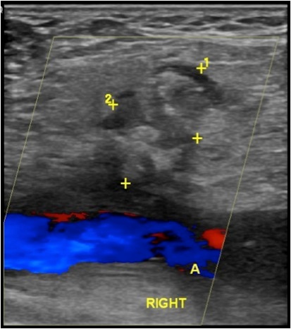
Figure 1 Ultrasound displaying a 2.1 x 1.4 x 1 cm heterogeneous area in the right common femoral artery demarcated by “+”.
Figure 1 Ultrasound displaying a 2.1 x 1.4 x 1 cm heterogeneous area in the right common femoral artery demarcated by “+”.
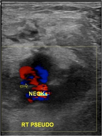
Figure 2 Repeat ultrasound displaying a pseudoaneurysm with the classic “neck”appearance depicted by the arrow.