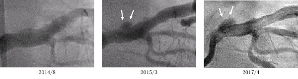
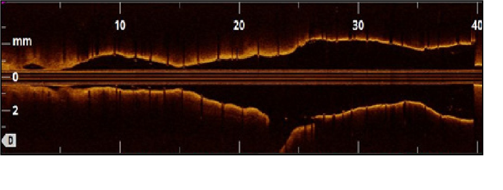
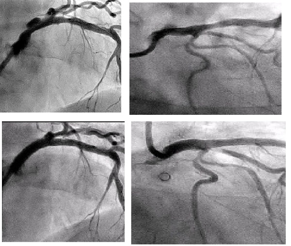
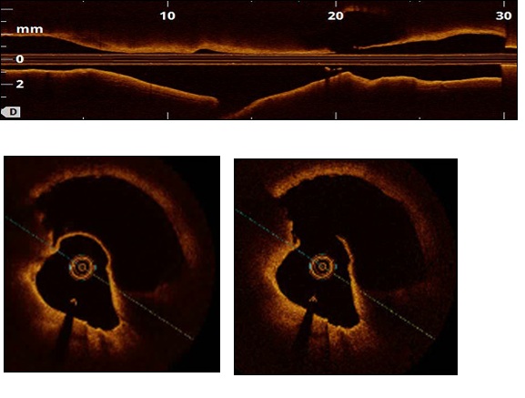
Figure 1A OCT revealed that vascular pseudoaneurysm neck had a lesion length of 6.6 mm, diameter of 2.7 mm, and width of 3.5 mm (including pseudoaneurysm 4.8×6.0 mm).
Figure 1B.1C OCT of the pseudoaneurysm showed a break in a part of the coronary artery vascular wall, with the site lacking all three layers (i.e., the tunica intima, media, and externa). It was considered to be a pseudoaneurysm.
Figure 2 The patient had undergone coronary intervention at another hospital on August 2014 and March 2015, and the present coronary angiography showed a clear trend of increasing size when compared to the sizes of the coronary artery pseudoaneurysm.
Figure 3 OCT was used to confirm resolution of the pseudoaneurysm and favourable stent bonding, and the GraftmasterŽ (Abbott Vascular., Co. Ltd., Tokyo, Japan) was shortened from the original 16 mm to approximately 13.3 mm so as not to protrude into the aorta.
Figure 4 Images before PCI (above) and after PCI(below). PCI utilizing the GraftmasterŽ (Abbott Vascular., Co. Ltd., Tokyo, Japan) enabled us to repair a coronary artery pseudoaneurysm.