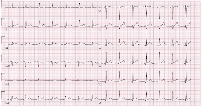
Figure 1A Electrocardiogram. Diffuse ST elevations seen in anteroseptal, lateral and inferior leads, and QTc prolongation.
Figure 1A Electrocardiogram. Diffuse ST elevations seen in anteroseptal, lateral and inferior leads, and QTc prolongation.
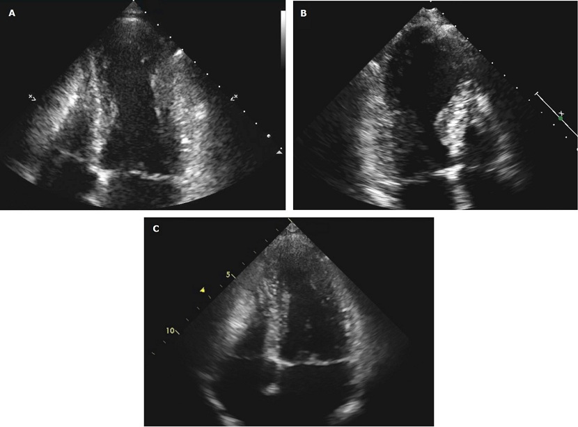
Figure 2 Transthoracic echocardiogram. A and B. Apical four chamber views demonstrating dilated akinetic apex and LV dysfunction typically seen in takotsubo cardiomyopathy. C. TTE repeated four months later with resolution of LV dysfunction.