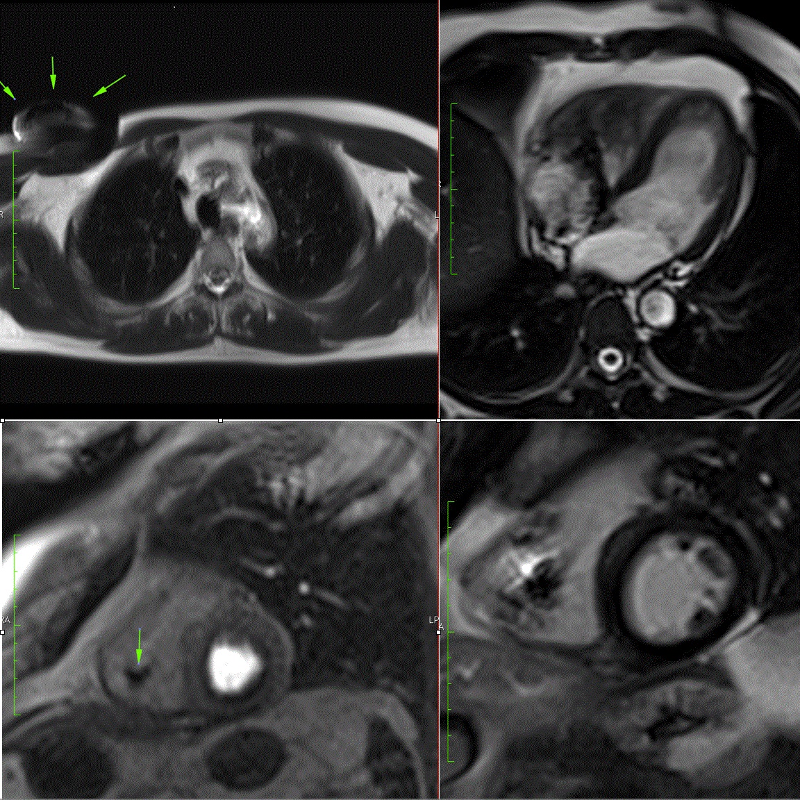
Figure 1
Left upper:Axial T2 weighted image showing the pacemaker (arrows) in the right subpectoral region
Right upper:SSFP cine image in 4-chamber view orientation in diastole
Left lower Perfusion image after 3 minutes of adenosine infusion with mild subendocardial perfusion defect in a non-ischemic distribution pattern, consistent with ring-artifact
Right lower Late Gadolinium Enhancement image in short axis orientation, showing normal myocardial appearance without relevant uptake