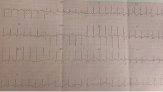
Figure 1 Electro cardiogram showing right
Figure 1 Electro cardiogram showing right
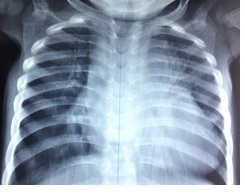
Figure 2Chest X-ray showing cardiomegaly at the Ventricular hypertrophy expense of right cavities and vascular redistribution to the apices.
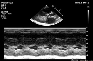
Figure 3M-mode para-sternal long axis view revealing
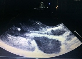
Figure 4shows concentric left ventricular hypertrophy and small aortic ring.
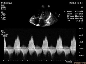
Figure 5Continuous wave Doppler on the mitral valve evaluating the elevation of the mitral gradient.