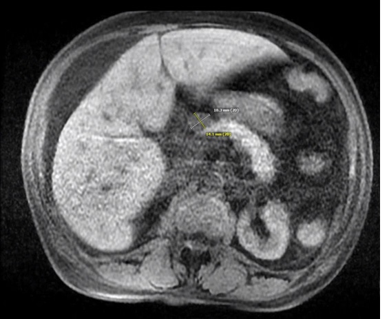
Figure 1 MRI demonstrating normal liver capsule with a 1.8 x 1.4 cm mass in the anterior aspect of the pancreatic body.
Figure 1 MRI demonstrating normal liver capsule with a 1.8 x 1.4 cm mass in the anterior aspect of the pancreatic body.
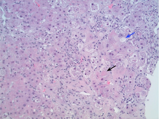
Figure 2 H&E stain at 200X magnification showing inflammation with Mallory's hyaline (black arrow) with few areas of balloon degeneration reflective of mild steatosis (blue arrow).
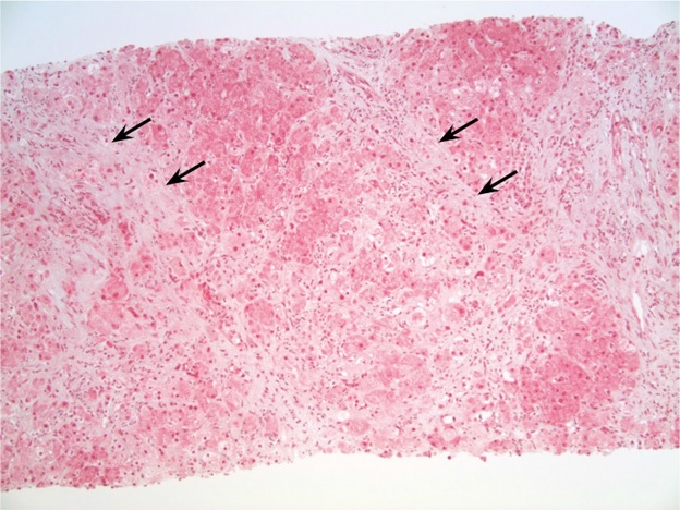
Figure 3 Trichrome stain at 100X magnification demonstrating fine chicken-wire fibrosis (black arrows) without cirrhosis or significant steatosis.