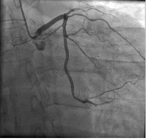
figure 1. LAO caudal projection shows the LAD artery was absent, the intermediate branch had 99% stenosis in the proximal segment
figure 1. LAO caudal projection shows the LAD artery was absent, the intermediate branch had 99% stenosis in the proximal segment
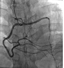
figure 2. RAO cranial view demonstrates the anomalous LAD originating from the proximal of RCA segment and had 80% stenosis in the distal segment
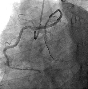
figure 3. The finally results of angiography after PCI procedure.
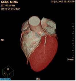
figure 4. Reconstructed three-dimensional image obtained by volume-rendering technique shows the anomalous LAD and coursed between the aorta and pulmonary artery
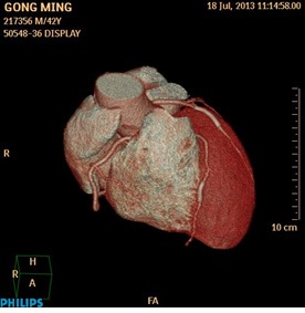
figure 5. Reconstructed three-dimensional image illustrates stent was implanted in the intermediate branch and the distal of LAD segment seperately.