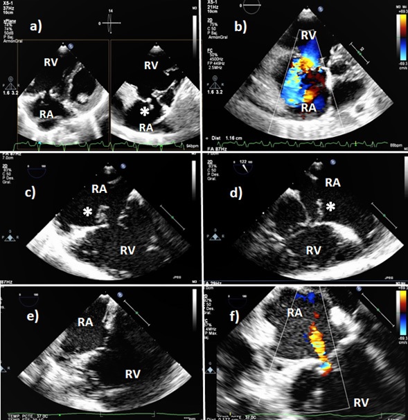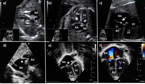
figure 1. A two-dimensional fetal echocardiogram (a-c) with localized areas of increased echogenicity at the papillary muscles of both AV valves. The last fetal echocardiogram (c) showed minimal residual echo bright density limited to the tricuspid valve (arrow). Postnatal echocardiogram showed the tricuspid valve with an image resembling vegetation (asterisk) and, by color-flow Doppler, severe insufficiency of the tricuspid valve (d-e). RV: right ventricle. RA: right atrium. LV: left ventricle. LA: left atrium.