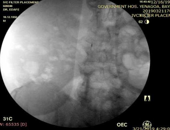
Figure 1: infra-renal IVC segment at L3
Figure 1: infra-renal IVC segment at L3
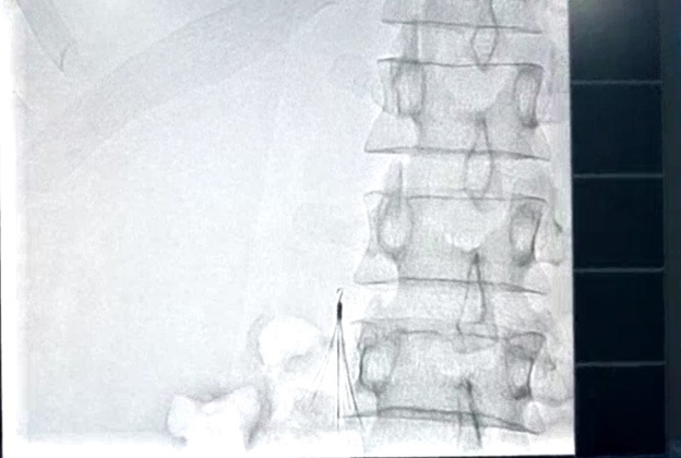
Figure 2: infra-renal ALN IVC filter.
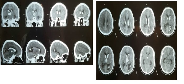
Figure 3: brain Computer Tomography showing intra-ventricular bleed
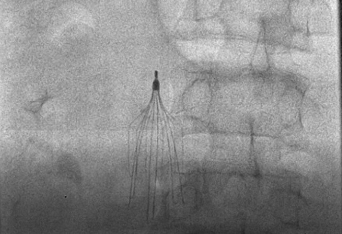
Figure 4: Infra-renal IVC filter (denali filter)
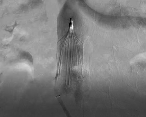
Figure 5: post-deployment venogram
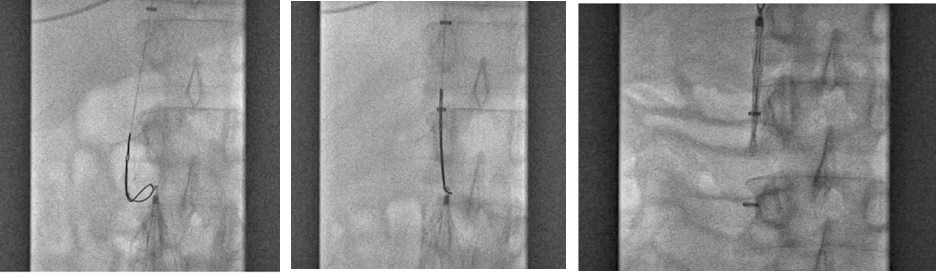
Figure 6. Retriever of the IVC filter
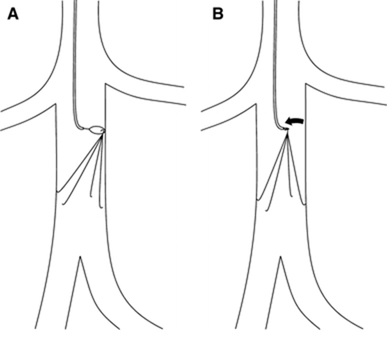
Figure 7: Realignment technique (loop snare with single access). A Curved guiding catheter is used to redirect the snare toward the filter apex. B Filter apex is grasped with the snare and realigned with the axis of the cava (curved arrow). This was the procedure done for case 3 patients.
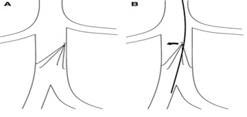
Figure 8: Stiff wire-displacement technique shown in A and B
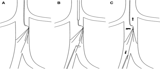
Figure 9: Dual-access technique (wire and snare with dual access). A Wire is introduced by way of jugular access and directed between filter apex and cava wall with the aid of a curved-tip guiding catheter. B The wire is snared by way of the femoral approach. C Wire traction is applied simultaneously in caudal and cephalic directions (straight arrows) resulting in the displacement of the filter apex from the cava wall (curved arrow).
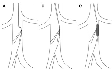
Figure 10: Balloon-Displacement Technique in A, B and C

Figure 11: Balloon-Displacement Technique in A, B and C
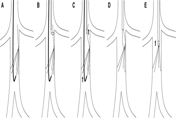
Figure 12: Sling Technique in A, B, C, D, and E
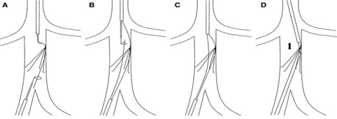
Figure 13: Parallel wire and dual-sheath technique in A, B, C and D
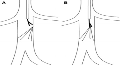
Figure 14. Dissection Technique