Global Research Trends in Cutaneous Neurofibromas: A Bibliometric Analysis from 2003 to 2022
Received Date: July 11, 2025 Accepted Date: July 26, 2025 Published Date: July 30, 2025
doi: 10.17303/jdct.2025.1.103
Citation: Jiani Wang, Jie Fu, Yu Zhou, Dongmei Gao, Jihong Qing, Guoke Yang (2025) Global Research Trends in Cutaneous Neurofibromas: A Bibliometric Analysis from 2003 to 2022. J Dermatol Cosmet Ther 1: 1-19
Abstract
Background: Neurofibromatosis type 1 (NF1) is a common inherited disorder characterized by cutaneous neurofibromas and other features. It is still a challenge in managing inoperable patients and the complex nature of the disease. Exist for Bibliometric analyses for cutaneous neurofibromas (cNF) could offer insights into impactful research and collaborations, guiding future efforts to improve patient care and outcomes.
Methods: We conducted a comprehensive literature search of the Web of Science Core Collection database for the period 2003-2022. Data processing and analysis were performed using bibliometric tools including VOSviewer, CiteSpace and “Bibliometrix” package. Our analysis assessed the publication or collaboration of countries, institutions, authors, and journals, as well as the co-citation and burst of references and keywords.
Results: The analysis included 927 articles from 465 journals and 1,402 institutions in 67 countries. Research on cNF has been increasing in recent years. The United States leads the field. Pierre Wolkenstein was the top author, while The University of Hamburg was the most productive institution. The American Journal of Medical Genetics Part A published the most articles in cNF. Co-citation analysis revealed major research topics and trends over time, showing growing interest in evaluating quality of life and genotype-pheno-type correlation for cNF patients. Emerging topitcal MEK inhibitors show potential as a promising therapy.
Conclusion: In conclusion, our bibliometric analysis of cNF research over the past two decades highlights the growing interest in this complex genetic disorder. Leading countries, authors, institutions, and journals have played significant roles in shaping the field. Notably, recent trends emphasize the importance of evaluating quality of life and genotype-phenotype correlations in cNF patients. Furthermore, the emergence of promising topical therapy marks an exciting development in the quest to improve patient care and outcomes for those affected by cNF, paving the way for future research and collaboration.
Keywords: Cutaneous Neurofibroma; Skin Benign Tumor; Bibliometircs; Vosviewer; Citespace
Introduction
Neurofibromatosis type 1 (NF1) is one of the most common inherited neurocutaneous disorders, affecting approximately 1 in 2,000 individuals [1]. Patients with NF1 commonly develop cutaneous neurofibromas, affecting up to 95% of patients [2,3]. Other characteristics include multiple café au lait spots, Lisch nodules, and axillary freckling. NF1 results from mutations in the NF1 gene on chromosome 17, which encodes neurofibromin. Loss of neurofi- bromin function activates the RAS signaling pathway, leading to overgrowth of cells [4,5]. Cutaneous neurofibromas (cNF) are benign tumors originating from peripheral nerves beneath the skin. They arise from abnormal proliferation of Schwann cells, which normally myelinate and support neurons. Mutations in the NF1 gene trigger this aberrant Schwann cell growth, forming neurofibromas [6].
cNF typically emerge during adolescence and continue to enlarge during a patient's lifetime [7]. While benign, they can cause pain, cosmetic issues, functional impairments and reduced quality of life [8,9]. Current treatments aim to manage symptoms but are often only partially effective. Some NF1 patients have too many, large, or difficult-- to-access neurofibromas, making surgical removal impossible and posing long-term management challenges [10]. Improving treatments and quality of life for NF1 patients, especially those with inoperable disease, remains an active area of research. However, the complexity and heterogeneity of cNF and difficulties managing inoperable patients mean that gaps in knowledge remain. map and analyze the scholarly literature, providing an objective perspective that complements traditional reviews [11]. By visualizing trends over time, they can clarify how knowledge has developed, identify emerging areas of high productivity, and highlight novel approaches and hypotheses. However, no bibliometric studies currently exist for cNF. For this disease, such analyses could reveal the most impactful researchers, institutions and countries, as well as major publishing journals. The study implications would suggest priorities to optimize and focus future research efforts on improving care and outcomes for patients.
Methods
Methodologies for Search and Collection of DataIn academic circles, the bibliometric analysis data extracted from WoS enjoys widespread credibility as the most reliable data source [12]. For the present study, we undertook a thorough literature search using the Web of Science Core Collection (WoSCC) database, encompassing the period from January 1, 2003, to December 31, 2022. To achieve a comprehensive and precise outcome, we deployed the following search strategy: ((TS=(cutaneous) OR TS=(- derma*) OR TS=(skin))) And ((TS=("neurofibromatosis type 1") OR TS=(“neurofibromatosis 1") OR TS=("von Recklinghausen’s disease") OR TS=("von Recklinghausen neuro- fibromatosis") OR TS=("von Recklinghausen’s phakomatosis") OR TS=("peripheral neurofibromatosis")))OR(TS=("cutaneous neurofibroma*" ) OR TS=("skin neurofibroma*" ) OR TS=("dermal neurofibroma*")). We confined our search to articles and reviews, while excluding other document types, such as meeting abstracts, letters, and editorial materials. Ultimately, our analysis comprised 927 records, and the particulars of the literature screening process are elaborated in Figure 1.
Data AnalysisInitially, the data sourced from WoSCC was imported into Microsoft Excel 2021.Subsequently, two authors carried out separate screening of the articles included in the final selection and extracted all relevant details pertaining to the included papers, which comprised titles, authors, keywords, institutions, countries/regions, citations, journals, and publication dates. The processed data was subsequently examined utilizing bibliometric tools, such as VOSviewer (version 1.6.19), CiteSpace (version 6.2.R2), and the R package "bibliometrix".
CiteSpace is a bibliometric tool that enables the exploration and visualization of prevailing trends and patterns within a given research sphere. Moreover, it produces a knowledge map of related domains, affording an extensive outlook on a specific field of expertise, and applies diverse dynamic network analysis methods to recognize crucial studies, leading-edge research, evolving developments, and frontiers within a particular scientific field [13-16]. In this investigation, CiteSpace was the tool of choice for conducting co-citation studies. The settings for CiteSpace were established as follows: in the Time Slicing column, the time span was delineated from January 2003 to December 2022, with each year represented as a separate time slice.
VOSviewer is a software instrument developed by the Leiden University Center for Science and Technology Studies that facilitates the generation and appraisal of bibliometric networks. this application exhibits the ability to construct bibliographic networks, specifically co-authorship, co-occurrence, and citation-based relationships in bibliographic data [17-19]. Additionally, co-occurrence and cluster analyses on authors, research institutions, countries, and discipline aspects were executed utilizing either CiteSpace or VOSviewer, as part of this study
The Bibliometrix software package, developed by Dr. Massimo Aria and Corrado Cuccurullo of Naples University in Italy, is an open-source tool that enables thorough analytical inquiry into bibliometrics and scientometrics [20]. In this study, the software was implemented to promote assessment of authors' H-index and publications evolution over time.
Results
Annual Publication TrendsThe overall trend of increasing published articles can be temporally bifurcated into two phases (Figure 2). The first phase spans from 2003 to 2014, during which the annual number of articles published, with the exception of 2010, registered less than
40. The majority of annual volumes fluctuated between 25-40. In contrast, the second phase encompasses the time period of 2015-2022, where the number of published articles ascended abruptly to 58 in the year 2015. Subsequently, the annual volumes of published articles remained higher than 40, peaking at 96 articles in 2021, which is 3.5 times more than that in 2003.
Research Contributions by CountriesTable 1 outlines the publication statistics for selected countries in the field. The United States holds the highest number of publications with 251, making up 27.08% of the total publications. Japan follows with 70 publications while Germany, China, and Italy have 69, 65, and 65 publications, respectively. The minimal difference in the numbers emphasizes the U.S.'s dominance in this field, as its total number of publications almost equals the sum of the remaining four countries' publications. Notably, the H-index is also highest for the U.S., underscoring its undeniable contribution and influence in the field. Examination of China, Turkey, India, and Brazil, who rank in the top 10 for publication count, reveals a relatively weak h-index and g-index, indicating poor overall quality or insuficient publication in prestigious journals. The findings suggest a need for these developing countries to enhance the quality of their research output.
For the foremost five nations, we present the annual variations in the publication count, as exemplified in Figure 3. The size and hue of the circles concomitantly mirror the number of publications. Notably, Germany's publication count has exhibited rather steady progression over the prior two decades, while the remaining nations display comparatively ascending tendencies in recent years. In particular, China's publication count has markedly exceeded previous figures over the last five years, thus suggesting that China shall contend a more prominent position in this domain going forward.
In this study, we utilized VOSviewer to visualize the inter-country cooperation network. As presented in Figure 4A, the countries with the most extensive inter- country cooperation were the United States (links=180), followed by Germany (links=111) and the United Kingdom (links=108). In contrast, India (links=8), China (links=4), Iran (links=2), South Korea (links=1), and other Asian regions demonstrated limited collaboration, indicating the need for these countries to enhance their cooperation with other nations to improve the generalizability of the study.
Furthermore, we employed the bibliometric.com website for online mapping, and it also demonstrated that European countries, including the United States, Germany, the United Kingdom, and France, reported more substantial cooperation (Figure 4B).
Productivity and Collaboration of Research InstitutionsTable 2 displays the leading 10 institutions arranged in a descending order of publication count, on the basis of the corresponding author's affiliation. Among the listed institutions, four are situated in the United States, namely the University of Texas Southwestern Medical Center (n=14), the University of Alabama at Birmingham (n=12), Johns Hopkins University (n=11) and Indiana University (n=10). This indicates that the United States is home to several top-tier research institutions, which contributes to its substantial national publication output. The University of Hamburg (n=24) and the University of Ulm (n=11) together contribute greater than half of the total number of publications generated in Germany, underscoring the concentration of German research centers in this area at these two institutions.
Notably, Shanghai Jiao Tong University is the sole Asian institution featured on the list; however, its citation rate and institutional H-index are below average, highlighting the need for it to prioritize improving article quality to gain recognition from other research institutions in the field in the future.
We imported the documents from the Web of Science Core Collection database into Vosviewer and subsequently identified 1402 institutions. Using a minimum publication count of 5, the list was narrowed down, including 71 institutions which met the criteria required for our analysis. Based on their level of collaborative interaction, these selected institutions were segregated into eight clusters (Fig 5A). Cluster 1 comprised mainly of Harvard University, Massachusetts General Hospital, and Indiana University. On the other hand, Cluster 2 included Paris-East Créteil University, Hôpital Henri-Mondor, and Paris Descartes University (now known as Université Paris Cité). Cluster 3 was primarily made up of University of Alabama at Birmingham, Johns Hopkins University, and Cardiff University, while the primary institutions in Cluster 4 consisted of University Medical Center Hamburg-Eppendorf, University of Hamburg, and University of Ulm. The primary institutions in Cluster 5 were Federal University of Rio de Janeiro, University of São Paulo, and Fluminense Federal University, while Sapienza University of Rome, University of Padua, and Catholic University of the Sacred Heart were the main institutions in Cluster 6.
Finally, Cluster 7 primarily included the Neurofi- bromatosis Institute (a non-profit organization in the United States), University of Florida, and Charles University in Prague. Cluster 8 predominantly constituted the University of Turku, Turku University Hospital, and University of Oulu. It is evident that institutions within the same country tend to coalesce within clusters, wherein inter-institutional collaborations are often limited to the same region or country, as opposed to international collaborations. Notably, the University of Alabama at Birmingham (links=29) in the United States exhibits the highest number of collaborative efforts with other institutions, while Paris-East Créteil University (links=22) in France demonstrates the most external collaborations in comparison to other French institutions. these can be considered as two representative research institutions within this field.
Analysis of Authorship on cNF ResearchAn analysis of the cNF research field was conducted using the "biliometrix" R package, with data sourced from the WOSCC database. The results reveal the top ten authors in the field, including Pierre Wolkenstein (n = 38), Victor-Felix Mautner (n = 30), Reinhard Friedrich (n = 25), Meena Upadhyaya (n = 20), Lan Kluwe (n = 19), Dominique Vidaud (n = 19), Hildegard Kehrer-Sawatzki (n = 16), Lu Q. Le (n = 15), Christian Hagel (n = 14), and Béatrice Parfait (n = 13). Further details regarding the number of published papers, total citations, and H-index for these authors are presented in Table 3. Notably, a significant proportion of the top authors (five out of ten) are affliated with the same institution, suggesting strong collaboration between research groups within the institution.the list of top experts comprises five medical geneticists (Meena Upadhyaya, Lan Kluwe, Dominique Vidaud, Hildegard KehrerSawatzki, and Béatrice Parfait), as well as dermatologists (Pierre Wolkenstein and Lu Q. Le), neurologists (Christian Hagel and Victor-Felix Mautner), and a maxillofacial surgery professor (Reinhard Friedrich). Collaborating among specialists from diverse fields facilitates a comprehensive understanding of the complex nature of the condition. this multidisciplinary approach is crucial for improved diagnosis and treatment of patients.
Author co-occurrence graphs were constructed using Vosviewer (Figure 5B). the analysis revealed that Pierre Wolkenstein (links=38) and Meena Upadhyaya (links=36) have engaged in collaborative work with researchers from diverse institutions, whereas other authors have displayed relatively constant collaborations, with greater cooperation present among authors from the same geographic regions or institutions. An example of this can be observed in Cluster 4 (yellow), where all authors from Germany within Table 3 (Victor-Felix Mautner, Reinhard Friedrich, Lan Kluwe, Hildegard Kehrer-Sawatzki, and Christian Hagel) are clustered.
In order to attain a more thorough comprehension of the authors' publishing output, a graphical representation was created to illustrate their annual publication volume and citation frequency progression over time (Figure6). As depicted in said graph, the majority of authors did not maintain a constant rate of article production from twenty years ago till the current time, and observed gaps of two to three years with no publications. Only Pierre Wolkenstein has consistently produced publications over the past five years. Although Lu Q. Le had an annual average of 2.5 relevant articles published during 2018-2021, he did not have any output in this field in 2022. We retrieved one publication on Malignant peripheral nerve sheath tumors (MPNSTs) authored by him. Béatrice Parfait and Lan Kluwe both have spans of four to five years during which no papers in this field were published, and they have not published many articles in recent years, indicating that their research focus may have shifted. However, Parfait B published several high-quality articles on SARS-CoV-2 in 2022, as well as an article on neurofibromatosis type 2. Similarly,
Journal Sources of Research OutputAmong the 465 journals included in the analysis, Table 4 presents the top ten in terms of publication volume, totaling 141 articles, which accounts for 15.21% of the analyzed articles. the American Journal of Medical Genetics Part A is the most widely read journal (n=25), followed by the Journal of Dermatology (n=19). Among the top journals, the Journal of Medical Genetics boasts the highest citation rate, averaging 73.64 article citations. the British Journal of Dermatology, the oficial publication of the British Association of Dermatologists, is a leading journal in the dermatology field, holding an exclusive position with an impact factor exceeding 10.
The dual-map overlay technique, which juxtaposes journals and disciplines, proves to be an effective method for illustrating the distribution of academic journals across diverse fields, tracing the development of citation pathways, and drawing attention to the evolving scientific research foci(25,26). To this end, we adopted citespace to perform a dual-map overlay analysis. Specifically, the left panel of the figure presents the distribution of citing journals, while the right panel depicts the distribution of cited journals. The lines connecting them signify their citation relationships (Figure 7). The analysis identified four main citation paths. Articles published in the molecular/biology/genetics disciplines were often cited by articles published in the molecular/biology/immunology, medicine/medical/clinical or dentistry/dermatology/surgery disciplines. However, Articles published in the health/nursing/medicine disciplines were mostly cited by articles published in medicine/medical/clinical disciplines.
Trends and Content of Cited LiteratureThe co-reference network affords an exposition of the interrelations among diverse references, which is predicated upon their co-citation frequency and similarity. By virtue of the clusters that emerge from this network, one can identify groups of references that exhibit similarity in topics or themes, and thereby reveal key research areas, in- fluential papers, and evolutionary trends within a given field or domain [23]. The clusters that derive from the CiteSpace software are visually represented in Figure 8A, where the top 12 clusters are depicted by using the log- likelihood ratio (LLR) algorithm. The image epitomizes a variety of convex hulls which correspond to distinct clusters, such as mek inhibitor (cluster #0), germline mutations (cluster #1), ras/mapk (cluster #2), patient-reported outcomes (cluster #3), phenotype-genotype correlations (cluster #4), tumor progression (cluster #5), genetic counseling (cluster #6), hras (cluster #7), noonan syndrome (cluster #8), melanocyte (cluster #9), shp-2 (cluster #10), and genetic modifiers (cluster #11). Every node represented on the map refers to a cited reference.
A chronologically ordered representation of the network may illustrate the temporal evolution of the cited works, including their appearance and disappearance. Such an approach could effectively disclose the historical development, emerge of trends and likely directions that future research may take in a certain field or domain. The position of each reference on the timeline relates directly to its date of citation, and the most recent ones are located to the right of the graphical display (Figure 8B). By scrutinizing the timeline, we can gain valuable insights into the temporal distribution of literature instances in each cluster. Specifically, we can observe that research on gene mutations initiated at the earliest timeline point (#1), followed by studies focusing on the ras (#7) and its pathway (#2). Notably, Mek, a crucial member of the extracellular signal-regulated kinase (ERK) pathway, is an attractive candidate for treating neurofibroma, as its therapeutic benefits gradually become apparent. Moreover, numerous mek inhibitors have been developed and numerous clinical studies have been conducted to this end.
In Figure 9, the top ten references that demonstrate notable bursts of citations are presented, which highlights their significance in the cNF-related research field. The line graph shows the crimson segment representing the period during which the literature receives the most frequent citations. The length of the crimson segment correlates with the duration when the literature experiences elevated citation rates. Moreover, we have compiled the top ten references by bursts and presented the information in Table 5.
Analysis of co-occurring KeywordsA total of 66 keywords were found to have a minimum of 4 occurrences out of 1842 keywords. Based on the collective frequency distribution of these 66 keywords, we classified them into 4 distinct clusters utilizing vosviewer (as depicted in Figure 10A). Cluster #1 (colored red) predominantly comprises neurofibromas, schwannomas, and MPNSTs; cluster #2 (colored green) mostly encompasses cutaneous neurofibromas, café-au-lait spots and ras/mapk pathways; cluster #3 (colored blue) primarily includes neurofibromatosis type 1, plexiform neurofibromas and mek inhibitors; and cluster #4 (colored yellow) mainly encompasses genotype- phenotype correlation, tuberous sclerosis and mosaicism. The identification of bursts is deemed as an indicator of the fluctuating trends within certain fields or emerging topics across different timelines. Figure 10B illustrates the top ten keywords with the most potent burst intensity. In this figure, "Year" denotes the year of the first emergence of the keyword, while "Begin" and "End" respectively denote the inaugural and the final years of the reference burst where the said keyword appeared. Based on the depicted figure, recent research hotspots highlight "quality of life" and "genotype phenotype correlation".
Discussion
This is the first bibliometric analysis of research on cNF, examining over 20 years of literature. There was an overall increase in the number of published articles over time, which can be divided into two phases. The first phase from 2003 to 2014 saw a modest number of publications annually, potentially due to a lack of disease awareness, limited funding and few technological advances.The second phase from 2015 to 2022 experienced a dramatic rise in publications, beginning with a surge to 58 articles in 2015. This increase can be attributed to advances in diagnostic and therapeutic technologies like next-generation sequencing and induced pluripotent stem cells that enabled deeper investigation of cNF [27-29].
The United States was the leading contributor with the most publications and highest h-index. Other top countries like China, Turkey, India and Brazil showed weaker h-indices and g-indices, indicating a need to improve research quality and target high-impact journals. By enhancing their output, these developing countries could foster international collaboration and accelerate progress in the field. The United States' dominance can be attributed to factors such as robust funding, well-established institutions, a skilled workforce, and prestigious journals. These elements create a supportive research environment enabling high- -quality, influential publications.
the results show that research on cNF is concentrated in a few top institutions, mainly in the U.S. and Germany. While some Asian institutions contribute, Western institutions dominate in terms of output and impact. However, Asia has a large patient population, so findings from Asian studies could be influential. Collaboration between Eastern and Western institutions can effectively advance the field by leveraging Asia's large sample sizes and Western research expertise. Building global research capacity could help progress knowledge on cNF.
Analysis of institutional collaboration networks suggests that collaborative research on cNF is often geographically centered. While some institutions like the University of Alabama at Birmingham and Paris-East Créteil University show high collaborative activity and international partnerships, overall cross-border collaborations appear limited relative to domestic collaborations. Institutions seeking to collaborate may consider these two institutions first, given their extensive networks. Fostering international research consortia and funding for multi-country partnerships could help strengthen global collaborative networks in this field.
The top authors publishing on cNT represent fields like medical genetics, dermatology, neurology and maxillofacial surgery, reflecting the multidisciplinary approach needed [30,31]. Actually,the Neurofibromatosis Therapeutic Acceleration Program (NTAP) based at Johns Hopkins University School of Medicine is such a collaborative network of research centers bringing together clinicians and scientists across specialties to provide comprehensive care for NF1 patients. Analysis of author co-occurrence networks suggests that while some top authors have extensive international networks, others tend to collaborate more within their country or institution. Supporting broader cooperative networks across borders could strengthen research progress.
The current study finds that while several journals publish on cNT, few top journals account for most publications. Dermatology or plastic surgery journals tend to have lower impact factors due to their focused scope. Research in these fields often produces incremental findings rather than breakthroughs. There may also be a lag in translating basic science into clinical advances. The American Journal of Medical Genetics Part A and the Journal of Dermatology published the most articles. Dual map analysis suggests that articles published in molecular, biology and genetics journals are cited more often, indicating broader reach and higher impact. Therefore, researchers in dermatology or plastic surgery seeking to increase impact could consider publishing in molecular, biology and genetics journals in addition to specialty journals.
The co-citation network and timeline analysis provide valuable insights for understanding the current state and future directions of research on cNF. Key topics, influential papers and evolving research trends are identified that characterize the progression of knowledge in this specialized field. The emergence of MEK inhibitors as a promising therapeutic strategy is also evident [32-34]. Selumetinib, as a representative of the MEK inhibitor, has been studied for the treatment of pediatric neurofibromas in several clinical trials [35-37]. Phase 1 trials in children with neurofibromatosis type 1 (NF1) and associated plexiform neurofibromas (pNFs) showed that selumetinib shrank most patients' tumors [33]. Phase 2 trials in children with NF1 and inoperable plexiform neurofibromas demonstrated that selumetinib could achieve tumor shrinkage and reduce pain in some patients [38]. In April 2020, selumetinib was approved by the US Food and Drug Administration (FDA) for the treatment of NF1 and plexiform neurofibromas, making it the first effective treatment for this condition. Ongoing research aims to optimize MEK inhibitor therapy to make it effective for more NF1 patients while minimizing adverse effects. Given the benign nature of cNF, the use of oral MEK inhibitors may increase the risk-benefit ratio, so the search for topical MEK inhibitors with fewer side effects has led to the development of NFX-179 topical gel, which has success fully completed its Phase 1/2 a trial [39]. A randomized, double-blind, controlled trial of NFX-179 is currently underway enrolling nearly 200 patients [40].
The keywords clustering indicates that research spans from characterizing various tumor phenotypes to investigating underlying genetic mechanisms and potential therapies. Studies of tumor phenotypes and molecular pathways aim to identify potential drug targets while clinical research evaluates treatment options. The identification of keyword bursts over time reveals emerging research hotspots. "Quality of life" has become an increasingly important topic in recent years, suggesting growing interest in evaluating and improving patients' psychosocial wellbeing and functioning. Yuichi Yoshida et al. surveyed a large group of neurofibromatosis type 1 patients with cNF to gain insight into their real-world experiences and perspectives [41]. The majority of patients had cNF in visible areas of the body, and both the number and visibility of these skin growths increased with age. The patients reported that cNF mainly impacted their mood, daily life and social life, with visibility having a more significant effect than just the total number of growths. Over 75% of the patients expressed dissatisfaction with the frequent need for repeat operations and the high regrowth rate of cNF after surgery or laser therapy.
While surgical excision remains the primary treatment modality for cNF, the need for repeated excisions and resultant postoperative scarring can cause considerable psychological distress for patients. This indicates a need to explore more effective oral or topical medications that may supplement or reduce the need for surgery. Maguiness et al. found that patients with a higher burden of cNF, especially more visible and facial lesions, experienced significantly poorer skin-specific quality of life as assessed by the Skindex questionnaire [42]. However, the researchers also acknowledged certain limitations of the Skindex as an evaluation tool for cNF, noting that Riccardi scores exhibited poor correlation with Skindex domains. This suggested that the Skindex instrument should be utilized independently, as it may fail to capture all relevant factors pertaining to cNF that influence patients' quality of life. Recently, P. Wolkenstein from Université Paris-Est Créteil built an new tool to evaluates patient’s quality of life [43]. The cNF-Skindex as-sesses quality of life (QoL) across three domains - functioning, emotions, and symptoms - filling a gap by providing a patient-reported outcome measure for cNF in neurofibromatosis type 1 patients. Patients with over 50 cNF had the most impaired QoL, highlighting the value of the cNF-Skindex in measuring the impact of cNF burden. Consequently, the cNF-Skindex can be useful in clinical practice to assess baseline QoL, treatment effects, and may also support reimbursement of cNF therapies by demonstrating improvements in quality of life.
Establishing genotype-phenotype correlations has several important implications [44]. It can enable earlier diagnosis for infants and children with mild mutations that do not initially meet diagnostic criteria, allowing for earlier treatment and management. It can help determine eligibility for emerging targeted therapies since some treatments may work best for patients with specific NF1 mutations, requiring a genotype-directed approach. Genotype data can also aid in stratifying patients in clinical trials based on their likely response to help reduce heterogeneity and improve trial outcomes. Certain NF1 mutations have been linked to more severe phenotypes, including whole gene deletions and mutations in specific domains of the NF1 protein [45]. In contrast, mutations affecting residue Arg1809 have been associated with a milder phenotype characterized by an absence of neurofibromas [46]. Filomena Napolitano provides valuable insights into the mutational spectrum and genotype-phenotype correlations in neurofibromatosis type 1 [47]. Frameshift mutations were found to be associated with a higher risk of learning disabilities compared to other mutation types. Large cohort studies are necessary to better distinguish the common cNF from other more aggressive neurofibromas, improving diagnosis and management of patients.
Conclusion
In summary, this bibliometric analysis provides novel insights into the landscape of cNF research over the past two decades. There has been a marked increase in research activity from 2015 onwards, driven by technological advancements and growing recognition of the condition. However, research is still concentrated in a few Western institutions, with the United States dominating. Greater international collaboration and research capacity building globally could help accelerate advancements. Analysis of influential references and keywords reveals the progression of knowledge from phenotype characterization to understanding molecular pathways and potential treatments. Ongoing efforts should aim to translate basic insights into effective topical MEK inhibitors to improve outcomes for patients with cNF. There is growing interest in evaluating and improving quality of life for cNF patients, highlighted by the development of the cNF-Skindex patient-reported outcome measure. Establishing genotype-phenotype correlations could enable earlier diagnosis, stratification of patients for targeted therapies, and improving clinical trial outcomes. Overall, this bibliometric analysis provides a comprehensive and objective view of the current state and future directions of cNF research.
- Uusitalo E, Leppävirta J, Koffert A, Suominen S, Vahtera J, Vahlberg T, et al. (2015) Incidence and mortality of neurofibromatosis: a total population study in Finland. J Invest Dermatol 135: 904-6.
- Bergqvist C, Servy A, Valeyrie-Allanore L, Ferkal S, Combemale P, Wolkenstein P, et al. (1966) Neurofibromatosis 1 French national guidelines based on an extensive literature review since 1966. Orphanet Journal of Rare Diseases 15: 37.
- Ge LL, Xing MY, Zhang HB, Li QF, Wang ZC (2022) Role of nerves in neurofibromatosis type 1-related nervous system tumors. Cell Oncol 45: 1137-53.
- Xu GF, O’Connell P, Viskochil D, Cawthon R, Robertson M, Culver M, et al. (1990) The neurofibromatosis type 1 gene encodes a protein related to GAP. Cell 62: 599-608.
- Viskochil D, Buchberg AM, Xu G, Cawthon RM, Stevens J, Wolff RK, et al. (1990) Deletions and a translocation interrupt a cloned gene at the neurofibromatosis type 1 locus. Cell 62: 187-92.
- Carroll SL, Ratner N (2008) How Does the Schwann Cell Lineage Form Tumors in NF1? Glia 56: 1590-605.
- Huson SM (1993) Neurofibromatosis: Phenotype, Natural History and Pathogenesis. Journal of Medical Genetics 30: 351
- Guiraud M, Bouroubi A, Beauchamp R, Bocquet A, Grégoire JM, Rauly-Lestienne I, et al. (2019) Cutaneous neurofibromas: patients’ medical burden, current management and therapeutic expectations: results from an online European patient community survey. Orphanet J Rare Dis 14: 286.
- Peltonen S, Jannic A, Wolkenstein P (2022) Treatment of cutaneous neurofibromas with carbon dioxide laser: Technique and patient experience. European Journal of Medical Genetics 65: 104386.
- Duong TA, Bastuji-Garin S, Valeyrie-Allanore L, Sbidian E, Ferkal S, Wolkenstein P (2011) Evolving Pattern with Age of Cutaneous Signs in Neurofibromatosis Type 1: A Cross- Sectional Study of 728 Patients. Dermatology 222: 269-73.
- Donthu N, Kumar S, Mukherjee D, Pandey N, Lim WM (2021) How to conduct a bibliometric analysis: An overview and guidelines. Journal of Business Research 133: 285-96.
- Birkle C, Pendlebury DA, Schnell J, Adams J (2020) Web of Science as a data source for research on scientific and scholarly activity. Quantitative Science Studies 1: 363-76.
- Yang D, Wu X, Liu J, Zhou J (2022) CiteSpace-based global science, technology, engineering, and mathematics education knowledge mapping analysis. Front Psychol 13: 1094959.
- Chen C (2006) CiteSpace II: Detecting and visualizing emerging trends and transient patterns in scientific literature. J Am Soc Inf Sci 57: 359-77.
- Chen C, Ibekwe-SanJuan F, Hou J (2010) The structure and dynamics of cocitation clusters: A multiple-perspective cocitation analysis. J Am Soc Inf Sci 61: 1386-409.
- Chen C, Song M (2019) Visualizing a field of research: A methodology of systematic scientometric reviews. PLOS ONE 14: e0223994.
- McAllister JT, Lennertz L, Atencio Mojica Z (2022) Mapping A Discipline: A Guide to Using VOSviewer for Bibliometric and Visual Analysis. Science & Technology Libraries 41: 319-48.
- Markscheffel B, Schröter F (2021) Comparison of two science mapping tools based on software technical evaluation and bibliometric case studies. COLLNET Journal of Scientometrics and Information Management 15: 365-96.
- Van Eck NJ, Waltman L (2010) Software survey: VOSviewer, a computer program for bibliometric mapping. Scientometrics 84: 523-38.
- Aria M, Cuccurullo C (2017) bibliometrix: An R-tool for comprehensive science mapping analysis. Journal of Informetrics 11: 959-75.
- Peyre M, Tran S, Parfait B, Bernat I, Bielle F, Kala-marides M (2023) Surgical Management of Peripheral Nerve Pathology in Patients With Neurofibromatosis Type 2. Neurosurgery 92: 317-28.
- Magallon-Lorenz M, Terribas E, Fernández M, Requena G, Rosas I, Mazuelas H, et al. A detailed landscape of genomic alterations in malignant peripheral nerve sheath tumor cell lines challenges the current MPNST diagnosis.
- Durier C, Ninove L, Lefebvre M, Radenne A, Desaint C, Ropers J, et al. (2022) Neutralizing antibodies against SARS-CoV-2 variants following mRNA booster vaccination in adults older than 65 years. Sci Rep 12: 20373.
- Molino D, Durier C, Radenne A, Desaint C, Ropers J, Courcier S, et al. (2022) A comparison of Sars-Cov-2 vaccine platforms: the CoviCompare project. Nat Med 28: 882-4.
- Chen C, Leydesdorff L (2014) Patterns of connections and movements in dual-map overlays: A new method of publication portfolio analysis. Journal of the Association for Information Science and Technology 65: 334-51.
- Chen C, Dubin R, Kim MC (2014) Emerging trends and new developments in regenerative medicine: a scientometric update (2000 – 2014). Expert Opinion on Biological Therapy 14: 1295-317.
- Anastasaki C, Woo AS, Messiaen LM, Gutmann DH (2015) Elucidating the impact of neurofibromatosis-1 germline mutations on neurofibromin function and dopaminebased learning. Hum Mol Genet 24: 3518-28.
- Pasmant E, Parfait B, Luscan A, Goussard P, Briand-- Suleau A, Laurendeau I, et al. (2015) Neurofibromatosis type 1 molecular diagnosis: what can NGS do for you when you have a large gene with loss of function mutations? Eur J Hum Genet 23: 596-601.
- Krauthammer M, Kong Y, Bacchiocchi A, Evans P, Pornputtapong N, Wu C, et al. (2015) Exome sequencing identifies recurrent mutations in NF1 and RASopathy genes in sun- exposed melanomas. Nature Genet 47: 996.
- Chamseddin BH, Hernandez L, Solorzano D, Vega J, Le LQ. Robust surgical approach for cutaneous neurofibroma in neurofibromatosis type 1. JCI Insight 4: e128881.
- Ortonne N, Wolkenstein P, Blakeley JO, Korf B, Plotkin SR, Riccardi VM, et al. (2018) Cutaneous neurofibromas: Current clinical and pathologic issues. Neurology 91: S5-13.
- Klesse LJ, Jordan JT, Radtke HB, Rosser T, Schorry E, Ullrich N, et al. (2020) The Use of MEK Inhibitors in Neurofi- bromatosis Type 1–Associated Tumors and Management of Toxicities. Oncologist 25: e1109-16
- Dombi E, Baldwin A, Marcus LJ, Fisher MJ, Weiss B, Kim A, et al. (2016) Activity of Selumetinib in Neurofibromatosis Type 1-Related Plexiform Neurofibromas. N Engl J Med 375: 2550-60.
- Jousma E, Rizvi TA, Wu J, Janhofer D, Dombi E, Dunn RS, et al. (2015) Preclinical assessments of the MEK inhibitor PD-0325901 in a mouse model of neurofibromatosis. type 1. Pediatric Blood & Cancer 62: 1709-16.
- AstraZeneca (2023) A Phase 1 Open Label Study to Assess the Safety, Tolerability, Pharmacokinetics and Clinical Efficacy of Selumetinib, a Selective Mitogen Activated Protein Kinase Kinase (MEK) 1 Inhibitor, in Chinese Paediatric and Adult Subjects With Neurofibromatosis Type 1 (NF1) and Inoperable Plexiform Neurofibromas (PN).
- AstraZeneca (2023) A Phase I, Single-Arm, Sequential Study to Evaluate the Effect of Food on the Gastrointestinal Tolerability and Pharmacokinetics of Selumetinib After Multiple Doses in Adolescent Children With Neurofibromatosis Type 1 Related Plexiform Neurofibromas.
- AstraZeneca (2023) A Phase 1 Open Label Study to Assess the Safety, Tolerability, Pharmacokinetics and Efficacy of Selumetinib, a Selective Mitogen Activated Protein Kinase Kinase(MEK)1 Inhibitor, in JapanesePaediatric Subjects With Neurofibromatosis Type 1 (NF1) and Inoperable and Symptomatic Plexiform Neurofibromas (PN).
- Gross AM, Wolters P, Baldwin A, Dombi E, Fisher MJ, Weiss BD, et al. (2023) SPRINT: Phase II study of the MEK 1/2 inhibitor selumetinib (AZD6244, ARRY-142886) in children with neurofibromatosis type 1 (NF1) and inoperable plexiform neurofibromas (PN). Journal of Clinical Oncology.
- NF lection (2022) Therapeutics, Inc. A Randomized Double-Blind, Vehicle-Controlled, Parallel Group Phase 2a Study to Determine Safety, Tolerability, Pharmacokinetics, and Pharmacodynamic Activity of NFX-179 Gel in Subjects With Cutaneous Neurofibromas.
- NF lection (2023) Therapeutics, Inc. A Randomized, Double-Blind, Vehicle-Controlled, Parallel Group Phase 2 Dose-Response Study to Determine Safety and Effectiveness of Two Concentrations of NFX-179 Gel in Subjects With Cutaneous Neurofibromas.
- Yoshida Y, Ehara Y, Koga M, Imafuku S (2022) Health-related quality of life in patients with neurofibromatosis 1 in Japan: A questionnaire survey using EQ-5D-5L. J Dermatol 49: 1228-32.
- Maguiness S, Berman Y, Rubin N, Dodds M, Plotkin SR, Wong C, et al. (2021) Measuring the Effect of Cutaneous Neurofibromas on Quality of Life in Neurofibromatosis Type 1. Neurology 97: S25-31.
- Fertitta L, Bergqvist C, Armand M, Moryousef S, Ferkal S, Jannic A, et al. (2022) Quality of life in neurofibromatosis 1: development and validation of a tool dedicated to cutaneous neurofibromas in adults. Journal of the European Academy of Dermatology and Venereology 36: 1359-66
- Kehrer-Sawatzki H, Cooper DN (2022) Challenges in the diagnosis of neurofibromatosis type 1 (NF1) in young children facilitated by means of revised diagnostic criteria including genetic testing for pathogenic NF1 gene variants. Hum Genet 141: 177-91.
- Bettegowda C, Upadhayaya M, Evans DG, Kim A, Mathios D, Hanemann CO, et al. (2021) Genotype-Phenotype Correlations in Neurofibromatosis and Their Potential Clinical Use. Neurology 97: S91-8.
- Pinna V, Lanari V, Daniele P, Consoli F, Agolini E, Margiotti K, et al. (2015) p.Arg1809Cys substitution in neuro- fibromin is associated with a distinctive NF1 phenotype without neurofibromas. Eur J Hum Genet 23: 1068-71.
- Napolitano F, Dell’Aquila M, Terracciano C, Franzese G, Gentile MT, Piluso G, et al. (2022) Genotype- -Phenotype Correlations in Neurofibromatosis Type 1: Identi- fication of Novel and Recurrent NF1 Gene Variants and Correlations with Neurocognitive Phenotype. Genes (Basel) 13: 1130.
FIGURE 1
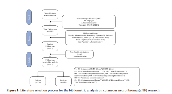
Figure 1: Literature selection process for the bibliometric analysis on cutaneous neurofibromas(cNF) research
FIGURE 2
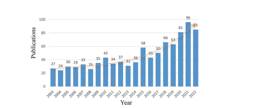
Figure 2: Annual publication trends on cNF research
FIGURE 3
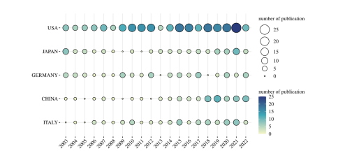
Figure 3: Publication output of the top 5 countries over time
FIGURE 4
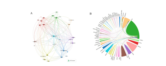
Figure 4: Country network analysis. (A) Network map of international collaboration among countries in cNF research (B) Country collaboration network in cNF research based on vosviewer
FIGURE 5
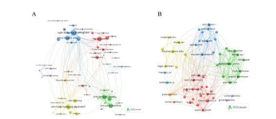
Figure 5: Institution co-occurrence analysis. (A) Collaboration network of institutions in cNF research; (B) Network visualization of author collaboration
FIGURE 6
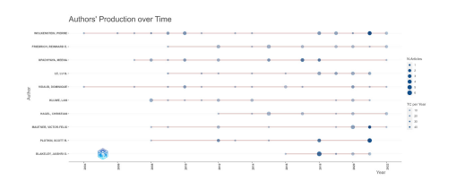
Figure 6: Publication trends of the top 5 highly productive authors
FIGURE 7
Figure 7: Dual-map overlay of citing and cited journals in cNF research
FIGURE 8
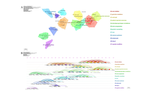
Figure 8: Co-citation analysis of references. (A) Clustered network map of co-cited references (B) Timeline visualization of co-cited references clusters
FIGURE 9
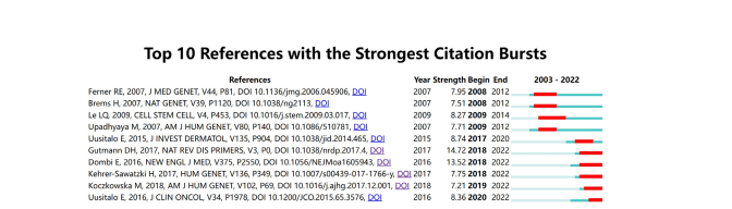
Figure 9: Top references with significant citation bursts. The red lines indicate the time period of the citation burst for each reference
FIGURE 10
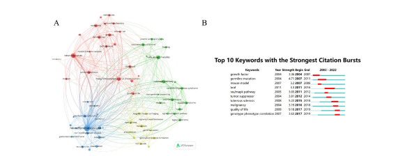
Figure 10: Keywords analysis (A) a network visualization of the keywords that most frequently co-occur in cNF research (B) Top 10 keywords that exhibit the strongest citation bursts in the literature on cNF
Tables at a glance
Figures at a glance