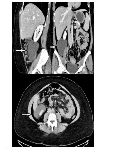
Figure 1: Computed Tomography demonstrating a well-defined fluid filling cystic lesion in the RLQ
Figure 1: Computed Tomography demonstrating a well-defined fluid filling cystic lesion in the RLQ
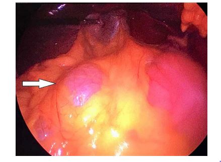
Figure 2: Intraoperative findings
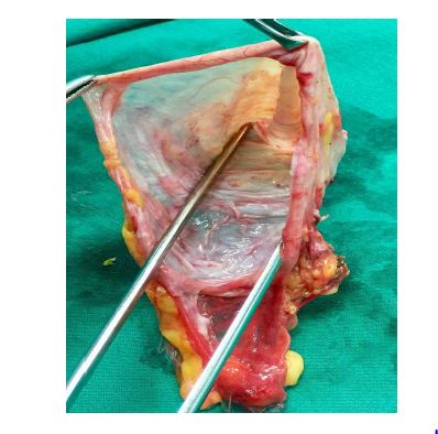
Figure 3: Gross Appearance of the cyst
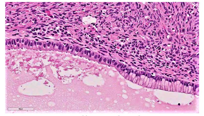
Figure 4: Histopathologic examination revealing mucinous lining
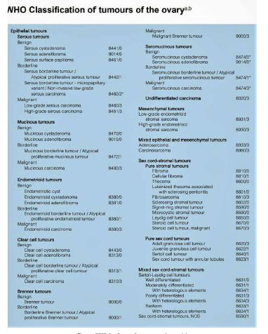
Figure 5: WHO classification of mucinous cystadenomas [6]
Tables at a glance
Figures at a glance