
Figure 1: Cross-sectional view of the CT abdomen and pelvis shows multiple dilated small bowel loop, with air fluid levels, a stenotic segment representing the transitional zone (red arrow) and fluid in the abdomen (yellow arrow)
Figure 1: Cross-sectional view of the CT abdomen and pelvis shows multiple dilated small bowel loop, with air fluid levels, a stenotic segment representing the transitional zone (red arrow) and fluid in the abdomen (yellow arrow)
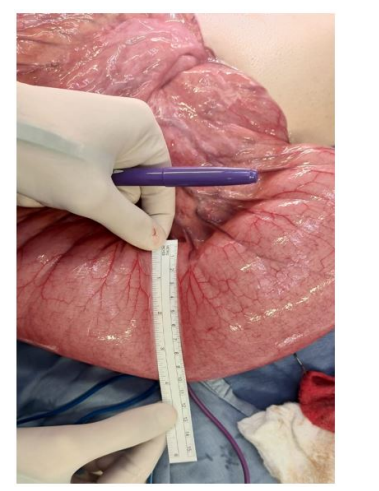
Figure 2: Intraoperative findings: remarkable small bowel distension reaching up to 10cm in diameter
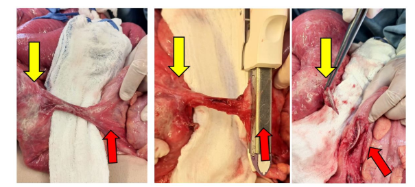
Figure 3: Intraoperative findings: fistulectomy at the sigmoidal end. Red arrow represents the sigmoidal end of the fistula and yellow arrow represents the ileal end
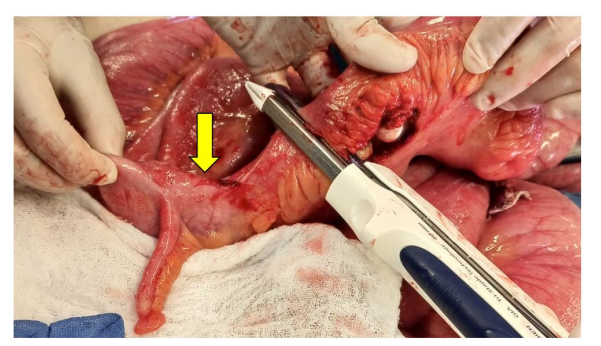
Figure 4: Intraoperative findings: enterectomy 3cm proximal to the ileocecal valve (yellow arrow)
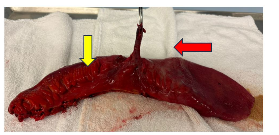
Figure 5: The resected ileal sigmoid which includes the stenosed ileum (yellow arrow) and fistula (red arrow)
Figures at a glance