Effect of Dentin Biomodification on Resin Dentin Bond Strength
Received Date: December 20, 2022 Accepted Date: January 20, 2023 Published Date: January 23, 2023
doi: 10.17303/jdoh.2023.10.101
Citation: Meenal Agarwal, Rajni Nagpal, Udai Pratap Singh, Chandrakar Chaman Mishra, Yashmin Parveen Karishma (2023) Effect of Dentin Biomodification on Resin Dentin Bond Strength. J Dent Oral Health 10: 1-11.
Abstract
Aim: To evaluate the effect of three different matrix metalloproteinase inhibitors on resin dentin shear bond strength.
Material and methods: Eighty-eight sound human molars extracted for periodontal reason were ground occlusally to obtain superficial dentin surfaces. Samples were allocated to following four groups: Group 1 no surface pretreatment was done; Group 2 Baicalein pretreatment; Group 3 Curcumin pretreatment and in Group 4 Berberine pretreatment was done before adhesive application. Resin composite restorations were placed after bonding procedures. Shear bond strength test was done at 24hrs (immediate) and at nine months (delayed). Interfacial adaptation was examined under scanning electron microscope. Bond strength values were statistically analyzed using two-way ANOVA and post hoc Tukey’s test at a significance level of p< .05.
Results: Delayed shear bond strength values of Baicalein and Curcumin groups were significantly higher than the control (p=0.001). However, Berberine treated group showed significantly lower bond strength than the control even at 9 months.
Conclusion Both baicalein and curcumin preserved the resin-dentin bond at 9 months’ storage period. However, berberine negatively influenced the bond strength both at immediate and delayed evaluation.
Keywords: Matrix Metalloproteinase Inhibitors; Resin-Dentin Bond; Baicalein; Curcumin; Berberine
Clinical Relevance: Incorporation of dentin biomodifiers into dental adhesives may impart MMP inhibitory potential and using these biofunctional adhesives may help in enhancing the stability of resin dentin interface.
Introduction
Stability of resin-dentin bond is a pre-requisite for the longevity of composite restorations. However, dental composites exhibit limited durability in vivo where resindentin bonded interface is the weakest link. Contemporary dental adhesives are simplified, user friendly systems which are water based and more hydrophilic than the conventional three-step adhesives. They allow water movement across the interface and do not provide a hermetic seal [1]. The integrity of the hybrid layer gets compromised due to the MMPs induced degradation of exposed collagen at the base of the hybrid layer as MMPs are hydrolases. MMPs are host derived enzymes, which play a role in degrading the extracellular matrix. MMPs remain inactive and entrapped in the calcified dentin matrix. They get activated by acidic agents used during bonding procedures. Along with this water induced adhesive degradation also occurs which reduces resin-dentin bond strength over time [2,3]. It has been reported that inhibition of MMPs may help in the preservation of resindentin bond. The deterioration of dentin bonding by endogenous MMPs can be prevented through application of dentin biomodifiers like MMP inhibitors or collagen crosslinkers which stabilize the dentin collagen. Crosslinking improves dentin mechanical properties and improves its resistance against enzymatic degradations [4,5]. Kiuru et al observed a lower loss of dentin bond strength with different MMP inhibitors and concluded that MMP inhibition can increase the longivity of resin dentin bond strength [6]. Yaghmoor et al also concluded that incorporation of MMP inhibitors into the adhesive system has a favourable effect on long term bond strength [7].
Various agents like chlorhexidine, glutaraldehyde, minocycline, proanthocyanidins, ascorbic acid, EDTA, carbodiimide, green tea extracts, quercetin, rosmarinic acid,and cashew nut shell liquid have been investigated to enhance resin dentin bond durability [8,9]. However, the effect of these agents on resin-dentin bond durability still needs to be clarified as there are variations in the concentration, type of solvent used and time of application of the biomodifying agents used in various studies. Use of natural collagen cross-linkers with MMP inhibitory potential might be advantageous as it induces low cytotoxicity, yetproviding stable resin–dentin bond.
Baicalein (BAI) is the major flavonoid found in Scutellaria baicalensis. BAI has been shown to inhibit MMP-2 and MMP-9 activity in tumor cells [10]. The inhibitory effect of BAI on these MMPs may be beneficial in dental adhesives [11].
Curcumin is a polyphenolic compound extracted from the plant Curcuma longa L. Curcumin, as isolated from the natural extracts, contains three major curcuminoids: curcumin, demethoxycurcumin, and bisdemethoxycurcumin. Curcuminoids have been extensively studied against various metabolic and infectious diseases [12-14]. The changes in the physico-chemical properties of collagen due to interaction with curcumin help in the accelerated wound healing process. Berberine, an isoquinoline alkaloid, can be isolated from Rhizoma coptidis, Hydrastis canadensis and Cortex phellodendri. Berberine has MMP inhibitory properties and has been shown to inhibit MMP 1, 2 and 9 in human gastric cancer cells [15]. However, the effect of these natural MMP inhibitors on resin-dentin bond durability has been controversially discussed in literature and no consensus has been reached [16,17]. Hence, the aim of this study was to investigate the effect of MMP-inhibitors Baicalein, Curcumin and Berberine on the preservation of dentin shear bond strength and also to observe micromorphology of the bonded interface under scanning electron microscope (SEM).
Materials and Methods
Eighty-eight extracted human molars were collected. The teeth free of caries, cracks, were included and examined under stereomicroscope (SZX10, Olympus, Tokyo,Japan). Teeth were cleaned and stored in 0.1% thymol solution for no more than 3 months. The teeth were collected after obtaining the patients inform consent under a protocol approved by institutional ethics and review board under protocol number KDCRC/IERB/11/2019/13.
Eighty tooth crowns were flattened occlusally using a low-speed diamond saw under water irrigation to expose superficial dentin. Teeth were placed in an auto polymerizing acrylic resin in such a way that only roots were vertically embedded while crown portions were above the resin.
Following solutions were prepared before starting with the adhesive procedures:
2.5μg/ml Baicalein solution: 2.5μg/ml Baicalein solution was prepared by dissolving it in 1% DMSO and 99% water.
50 μM Curcumin solution: 50 μM Curcumin solution was prepared by dissolving it in 1% DMSO and 99% water.
0.4% Berberine solution: 0.4% Berberine solution was prepared by dissolving it in 1% DMSO and 99% waters
Teeth were randomly divided into four groups (n =20) according to the dentin pretreatments. Shear bond strength for each group was evaluated at 24 hours (immediate) (I) and at 9 months (delayed) (D).
In all the groups, dentin surface was etched with etchant Detrey Conditioner 36 (Dentsply sirona) for 15 seconds, rinsed with water for 10 seconds and blot dried before application of MMP inhibitors.
Group 1: No Pretreatment: Acid etching with 35% phosphoric acid for 15 sec followed by application of distilled water for 1 min and gentle blot dried before application of etch and rinse adhesive Adper Single Bond 2 according to manufacturer’s instructions.
Group 2: Baicalein+DMSO: Application of 2.5μg/ml baicalein (Sigma Aldrich) dissolved in 1% DMSO on acid etched dentin surface for 1 min and gentle blot dried before application of the adhesive, Adper Single Bond 2 according to manufacturer’s instructions.
Group 3: Curcumin +DMSO: Application of 50μM curcumin (Sigma Aldrich) dissolved in 1% DMSO on acid etched dentin surface for 1 min and gentle blot dried before application of the adhesive, Adper Single Bond 2 according to manufacturer’s instructions.
Group 4: Berberine+DMSO: Application of 0.4% berberine (Vital herbs) dissolved in 1% DMSO on acid etched dentin surface for 1 min and gentle blot dried before application of the adhesive, Adper Single Bond 2 according to manufacturer’s instructions.
Transparent plastic tube with 3 mm internal diameter and 2 mm in height and thickness of 0.5 mm was placed at the center of the dentin surface in all the samples. The tubes were then filled with a nanohybrid resin composite Filtek Z250 XT (3M ESPE Dental Products) followed by light-curing for 20 seconds using 3M Elipar LED curing light.The plastic tubes were then cut and removed. All the restorations were completed by a single operator.
Ten samples from each group were stored in distilled water at 37°C for 24 hours before immediate shear bond strength testing and SEM analysis. The other ten samples from each group were stored in artificial saliva (Wet Mouth, ICPA Heath Products Ltd.) at 37°C in an incubator for 9 months before shear bond strength and SEM testing.
The specimens were placed in a universal testing machine (Instron, ADMET, Enkay Enterprises, New Delhi) and straight knife-edge rod (2.0mm) with a cross-head speed of 1mm/min was applied at the tooth restoration interface. Shear bond strength (MPa) was calculated by dividing the load at failure by surface area of the specimen. After bond strength testing, fractured samples were examined under stereomicroscope at 10 X magnification to differentiate failure pattern as Adhesive (a), Cohesive (c) & Mixed (m).
Scanning Electron Microscope Study
Two samples from each group were sectioned through coronal portion so as to obtain flat dentin blocks and subjected to treatments according to different groups followed by composite restoration till the height of 1mm. These were then sectioned bucco-lingually to expose the resin dentin interface and polished with (Shofu Super Snap Rainbow Technique Kit Ca) a series of increasingly finer SiC abrasive papers up to 1,200 grit and highly polished with a diamond paste.
The specimens were fixed in 10% formalin for 24 hours and subjected to acid-base treatment. Specimens were immersed in 6N HCl for 30 seconds, rinsed in distilled water followed by 10-minute immersion in 1% NaOCl, and final rinse with distilled water. The specimens were then dehydrated in ascending grades of ethanol upto 100% and kept in critical point dryer for 30 minutes. The specimens were gold sputtered and analyzed in scanning electron microscope.
Statistical Analysis
Values obtained from the shear bond strength were subjected to statistical analysis using SPSS (Statistical Package for Social Sciences) Version 21.0 statistical analysis software. Level of significance was p ≤0.05. The statistical tools used were Tukey’s HSD test, one-way ANOVA (multivariate assessment), Independent-t test and x2 test.
Results
Baicalein treated group (Group 2) depicted the highest immediate bond strength which was significantly higher than the control. Berberine treated group (Group 4) showed significantly lower bond strength than the control during immediate shear bond strength testing. When compared to the control group, the shear bond strength values of Baicalein and Curcumin treated group were significantly higher at nine months (p=0.001). However, Berberine treated group showed significantly lower bond strength than the control even at 9 months (Table 1). In SEM photomicrographs (Figure 1-4) Baicalein and Curcumin treated groups showed a good interfacial adaptation both at 24 hours and at nine months. However, interfacial gap was observed in the control group at 9 months and in Berberine treated groups both at 24 hrs and at 9 months.
Failure mode analysis: The distribution of failure pattern was compared using the chi square test (Figure 5). All the groups depicted mixed failures at 24 hours’ evaluation. While more adhesive failures were evident in all the groups after nine months with maximum number of adhesive failures in the Berberine treated group.
Discussion
Acid-etch procedure not only creates microporosities to be subsequently filled by adhesive resin, but also results in activation of dentinal matrix metalloproteases (MMPs), which degrade exposed collagen at the base of hybrid layer [18]. This deterioration of the interfacial seal leads to poor durability of composite restorations [16].
Many MMP inhibitors and collagen cross linkers have been studied for its use in adhesive dentistry like glutaraldehyde which is a widely known cross linker but its cytotoxicity limits its clinical use, proanthocyanidins caused discoloration of the treated dentin surface, riboflavin needs an additional light curing step reducing its clinical acceptability [19,20]. Use of natural MMP-inhibitors like EGCG, quercetin, baicalein, curcumin and berberine may be advantageous as compared to synthetic inhibitors and needs to be investigated as their effect may vary with varying concentration, time or adhesive system tested [21].
In this study, the results revealed that baicalein treated group showed significantly higher bond strength values after 24 hrs and at 9 months as compared to control and other experimental groups. These results indirectly support the collagen stabilization ability of BAI. BAI is a plant polyphenol similar to proanthocyanidins [22]. In accordance with our study, Zheng et al also showed that BAI was more effective in improving the enzymatic resistance of the dentin collagen matrix as compared to proanthocyanidins and quercetin [23]. They stated that due to its potential MMP inhibition and cross-linking properties, BAI could stabilize collagen fibrils and protect the integrity of the hybrid layer. In agreement with our results, Yi et al proved that ethanol wet bonding and baicalein enhanced the durability of resin - dentin bond [24]. In consensus with our study Li et al reported that Baicalein in a concentration of 2.5 μg/mL effectively increased microtensile bonding strength and resin-dentin bond durability and in a concentration range of 0-5.0 μg/mL it did not influence the conversion of adhesives [11].
MMP inhibitory mechanism of BAI may be attributed to various reasons: BAI could compete with the active center of the enzyme or cross-link and alter the threedimensional structure of MMPs [12]. It also might cross-link with dentin collagen fibers, changing the recognition sites of MMPs in collagen, thereby preventing their complexation with MMPs and resulting in the loss of enzymatic activity [11]. The hydrophobic nature of BAI may reduce water absorption thereby decreasing hydrolytic breakdown of collagen. Due to the polyphenolic structure of BAI it can directly scavenge free radicals. Chu et al explored the effect of various concentrations of BAI on the viability and cell cycle arrest of human dental pulp cells (HDPCs) and on the expression of matrix metalloproteinases (MMPs) and cathepsins in HDPCs [25]. They demonstrated that BAI at concentrations below 25 μmol/l did not affect cell viability in HDPCs while effectively inhibiting the expression of MMP-2, MMP-9, cathepsin-B, and cathepsin-K in HDPCs and improved the strength of aged resin-dentin bonding. They used baicalein in three different concentrations with 12.5 μmol/l showing the highest resin-dentin bond strength [25].
Baicalein could also inhibit the formation of S.mutans biofilm, thereby preventing restoration failure caused by secondary caries [26]. Chen et al depicted that the baicalein group substantially reduced enamel demineralization under biofilms and concluded that novel baicalein method is promising to inhibit dental caries by reducing biofilm formation and protecting enamel hardness [27].
Curcumin treated group depicted significantly higher shear bond strength values than control group at 9 months demonstrating that curcumin also preserved resindentin bond. This may be attributed to MMP inhibition ability of Curcumin as it chelates catalytic Zn2+ ions in MMPs and also causes crosslinking of the collagen fibrils thereby decreasing enzymatic degradation. Moreover, positively charged Curcumin has affinity for negative charges on the active sites of MMPs.12 Increase in tensile strength was observed in curcumin treated collagen, which suggests new crosslinking in collagen.13 Our results are supported by the study of Seseogullari et al who evaluated the endogenous MMP activity of demineralized dentin matrix following 1 and 5 min pretreatment by 20μM curcumin and 200 μM and found a significant reduction in MMP activity after 1 min of crosslinking treatment with curcumin [28]. Seseogullari et al in a subsequent research evaluated the effect of 50 or 100 μM curcuminoids on the dentin endogenous protease activity and reported decreased endogenous protease activity-mediated degradation in dentin [29]. 50–100 μM curcuminoids inhibited rhMMP-9 activity up to 50%. Similar to our study, Atay et al reported that after twelve months, bond strength of curcuminoids group was significantly higher than the controls and suggested the potential use of curcuminoids as protease inhibitors [30].
Curcumin is also being investigated as a root canal irrigant [14,31,32,33]. However, it is well known that turmeric concentrations have been correlated with increase in the reddish-yellow staining of nanohybrid composite resins, therefore use of curcumin may pose an aesthetic challenge.
Berberine treated groups demonstrated very low bond strength in both immediate and delayed testing.Although Tu et al proposed that berberine may slow periodontal degradation in periodontitis through regulation of MMPs and can in vitro effectively suppress the induced MMP-9 expressions in the induced macrophages but berberine seems to lack short term inhibition against endogenous dentin proteases [15]. Further studies are needed to assess the interactions of berberine with endogenous dentinal MMPs and methods for clinical application.
There were certain limitations of the study like lack of intrapulpal pressure during bonding, caries affected dentin was not studied and MMP inhibition was not directly evaluated. However, higher bond strength values at nine months in baicalein and curcumin groups suggest probable collagen stabilization which resulted in improved resindentin bond durability. Also, storing extracted teeth in thymol does not seem to have influenced dentinal MMP activity as reported in previous studies [34,35]. DMSO was used as a solvent for bio-modifiers in all experimental groups for standardization. Since DMSO was used with all agents in the current study, so relative comparisons could still be made amongst various biomodifiers notwithstanding that DMSO itself enhances dentin bonding [36].
Conclusion
Baicalein treated group showed the highest immediate and delayed bond strength values than other experimental groups. Both baicalein and curcumin preserved the resin-dentin bond at 9 months’ storage period. However,berberine negatively influenced the bond strength both at immediate and delayed evaluation. Further research is needed to determine a suitable clinical concentration and to explore the possibility of adding baicalein into dental adhesive to improve the stability of hybrid layer and maintain the durability of resin dentin bond in long term.
- Liu Y, Tjäderhane L, Breschi L, Mazzoni A, Li N et al. (2011) Limitations in bonding to dentin and experimental strategies to prevent bond degradation. J. Dent. Res 90:953-68.
- Betancourt DE, Baldion PA, Castellanos JE (2019) Resin-dentin bonding interface: Mechanisms of degradation and strategies for stabilization of the hybrid layer. Int J Biomater.
- Anumula L, Ramesh S, Kolaparthi VSK, Kirubakaran R, Karobari (2022) Role of Natural Cross Linkers in Resin–Dentin Bond Durability: A Systematic Review and Meta-Analysis. Materials 15: 5650.
- Abu Nawareg, Manar & Abuelenain, Dalia & Elkassas (2017) Role of dentin cross-linking agents in optimizing dentin bond durability. International Journal of Adhesion and Adhesives 78.
- Manuja N, Nagpal R, Pandit IK (2012) Dental adhesion: mechanism, techniques and durability. J Clin Pediatr Dent 36: 223-34.
- Kiuru O, Sinervo J, Vähänikkilä H, Anttonen V,Tjäderhane L (2021) MMP Inhibitors and Dentin Bonding: Systematic Review and Meta-Analysis. Int J Dent 9949699.
- Yaghmoor RB, Jamal H, Abed H, Allan E, Ashley P et al. (2022) Incorporation of MMP inhibitors into dental adhesive systems and bond strength of coronal composite restorations: A systematic review and meta-analysis of in vitro studies. Jpn Dent Sci Rev 58: 298-315.
- Hardan L, Daood U, Bourgi R, Cuevas-Suárez CE, Devoto W et al. (2022) Effect of Collagen Crosslinkers on Dentin Bond Strength of Adhesive Systems: A Systematic Review and Meta-Analysis. Cells 11: 2417.
- De Moraes IQS, do Nascimento TG, da Silva AT, de Lira LMSS, Parolia A (2020) Porto ICCM. Inhibition of matrix metalloproteinases: a troubleshooting for dentin adhesion. Restor Dent Endod 45: e31.
- Yan X, Rui X, Zhang K (2015) Baicalein inhibits the invasion of gastric cancer cells by suppressing the activity of the p38 signaling pathway. Oncol Rep 33: 737-43.
- Li J, Chen B, Hong N, Wu S, Li Y (2018) Effect of baicalein on matrix metalloproteinase and durability of resindentin bonding. Oper Dent 43: 426-36.
- Fathima NN, Devi RS, Rekha KB, Dhathathreyan A (2009) Collagen-curcumin interaction A physico-chemical study. J Chem Sci 121: 509-14.
- Panchatcharam M, Miriyala S, Gayathri VS, Suguna L (2006) Curcumin improves wound healing by modulating collagen and decreasing reactive oxygen species. Mol Cell Biochem 290: 87-96.
- Neelakantan P, Cheng CQ, Ravichandran V (2015) Photoactivation of curcumin and sodium hypochlorite to enhance antibiofilm efficacy in root canal dentin. Photodiagnosis Photodyn Ther 12: 108-14.
- Tu HP, Fu MM, Kuo PJ, Chin YT, Chiang CY et al. (2013) Berberine's effect on periodontal tissue degradation by matrix metalloproteinases: an in vitro and in vivo experiment. Phytomed 20: 1203-10.
- Cardoso MV, Mine A, Coutinho E, Van LK, De MJ et al. (2011) Current aspects on bonding effectiveness and stability in adhesive dentistry. J Aust. Dent 56: s31-44.
- Silva JC, Cetira Filho EL, Silva PGB, Costa FWG, Saboia VPA (2022) Is dentin biomodification with collagen cross-linking agents effective for improving dentin adhesion.A systematic review and meta-analysis. Restor Dent Endod 47: e23.
- Vidal CMP, Tjäderhane L, Scaffa PM, Tersariol IL, Pashley D et al. (2014) Abundance of MMPs and cysteine cathepsins in caries-affected dentin. J Dent Res 93: 269-74
- Hass V, Luque IV, Gutierrez MF, Moreira CG, Gotti VB et al. (2016) Collagen cross-linkers on dentin bonding:Stability of the adhesive interfaces, degree of conversion of the adhesive, cytotoxicity and in situ MMP inhibition. Dent Mater 32: 732-41.
- Fawzy AS, Nitisusanta LI, Iqbal K, Daood U, Neo J (2012) Riboflavin as a dentin crosslinking agent: ultraviolet a versus blue light. Dent Mater 28: 1284-91.
- Palma-Dibb RG, Roselino L De M, Neto PT, Faraoni JJ (2017) Strategies to Inactivate the Endogenous Dentin Proteases to Promote Resin-Dentin Bond Longevity in Adhesive Dentistry: A Critical Review. Reviews of Adhesion and Adhesives 5: 391-413.
- Chang W, Shao Z, Yin J, Chien C, Becker LB, Yuan C et al. (2007) Comparative effects of flavonoids on oxidant scavenging and ischemia-reperfusion injury in cardiomyocytes. J Eur Pharmacol 566: 58-66.
- Zheng K, Wu S, Chen B, Liao W, Li Y (2014) Effect of baicalein and quercetin on enzymatic resistance of dentin collagen. Zhonghua Kou Qiang Yi Xue Za Zhi 49: 667-71.
- Yi L, Yu J, Han L, Li L, Yang H (2019) Combination of baicalein and ethanol-wet-bonding improves dentin bonding durability. J Dent 90: 103207.
- Chu P, Li J, Lioa W, Wu S (2019) Effects of Baicalein on the Expression of Collagenolytic Enzymes in Human Dental Pulp Cells and Durability of Resin-Dentin Bonding. J Adhes Dent 21: 273-80.
- Vijayakumar A, Sarveswari HB, Vasudevan S, Shanmugam K, Solomon AP et al. (2021) Baicalein Inhibits Streptococcus mutans Biofilms and Dental Caries-Related Virulence Phenotypes. Antibiotics (Basel) 10: 215.
- Chen H, Xie S, Gao J, He L, Luo W et al. (2022) Flavonoid Baicalein Suppresses Oral Biofilms and Protects Enamel Hardness to Combat Dental Caries. Int. J. Mol. Sci 23: 10593.
- Seseogullari-Dirihan R, Apollonio F, Mazzoni A, TjäderhaneL, Pashley DH et al. (2016) Use of crosslinkers to inactivate dentin MMPs. Dent Mater 32: 423-32.
- Seseogullari-Dirihan R, Tekbas Atay M, Pashley DH, Tezvergil-Mutluay A (2018) Inhibitory effect of curcuminoid pretreatments on endogenous dentin proteases. Dent Mater J 37: 445-52.
- Tekbas Atay M, Seseogullari-Dirihan R, Mutluay MM, Tezvergil-Mutluay A (2022) Long-term effect of curcuminoid treatment on resin-to-dentin bond strength. Eur J Oral Sci 130: e12837.
- Frota MF, Guerreiro-Tanomaru JM, Tanomaru- Filho M (2015) Photodynamic therapy in root canals contaminated with Enterococcus faecalis using curcumin as photosensitizer. Lasers Med Sci 30: 1867-72.
- Sotomil JM, Münchow EA, Pankajakshan D (2019) Curcumin-A Natural Medicament for Root Canal Disinfection: Effects of Irrigation, Drug Release, and Photoactivation. J Endod 45: 1371-7.
- Tyagi P, Singh M, Kumari H (2015) Bactericidal activity of curcumin is associated with damaging of bacterial membrane. PLoS One 26: e0121313.
- Alharbi A, Saunders W, Jones S (2020) A new method for dentine matrix metalloproteinase extraction. Arch Oral Biol 113: 104694.
- Osorio R, Yamauti M, Osorio E, Ruiz-Requena ME, Pashley D et al. (2011) Effect of dentin etching and chlorhexidine application on metalloproteinase-mediated collagen degradation. Eur J Oral Sci 119: 79-85.
- Stape THS, Mutluay MM, Tjäderhane L, Uurasjärvi E, Koistinen A et al. (2021) The pursuit of resin-dentin bond durability: Simultaneous enhancement of collagen structure and polymer network formation in hybrid layers. Dent Mater 37: 1083-95.
FIGURE 1
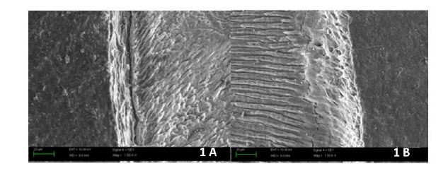
Figure 1: SEM photomicrograph of resin- dentin interface of Group 1 (No Pretreatment): Immediate (1A), Delayed (1B); interfacial gap is evident at nine months.
FIGURE 2
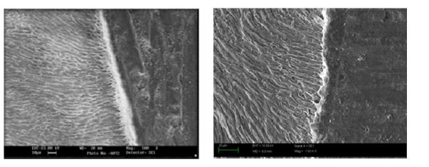
Figure 2: SEM photomicrograph of resin- dentin interface of Group 2 (Baicalein): Immediate (2A), Delayed (2B); good interfacial adaptation can be observed at both time periods.
FIGURE 3
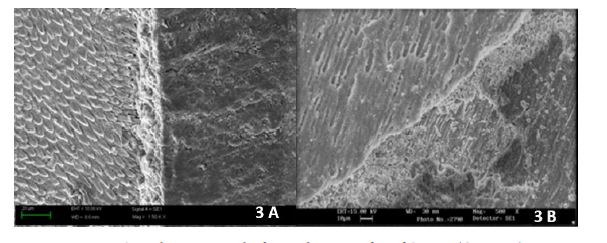
Figure 3: SEM photomicrograph of resin- dentin interface of Group 3 (Curcumin): Immediate (3A), Delayed (3B); good interfacial seal is evident.
FIGURE 4
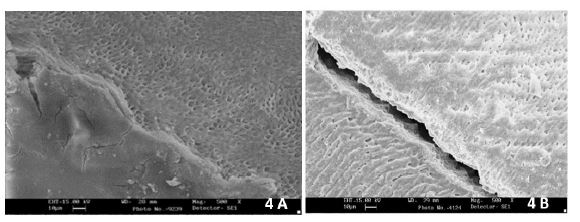
Figure 4: SEM photomicrograph of resin- dentin interface of Group 4 (Berberine): Immediate (4A), Delayed (4B); interfacial gap is observed at nine months.
FIGURE 5
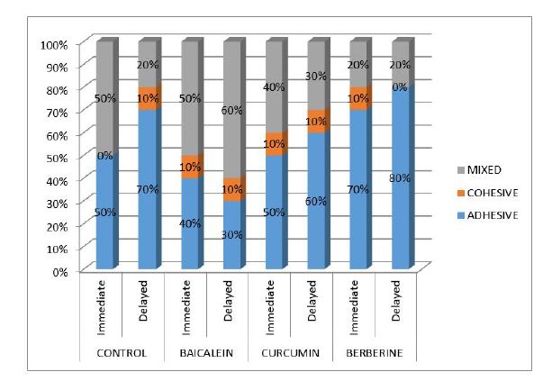
Figure 5: Failure mode analysis
Tables at a glance
Figures at a glance