Bifurcated Mandibular Second Premolars: Report of Unusual Root Anomaly (Supernumerary Root) – A Case Series
Received Date: May 23, 2023 Accepted Date: June 23, 2023 Published Date: June 26, 2023
doi: 10.17303/jdoh.2023.10.104
Citation: Nagaveni NB, Ashwini KS (2023) Bifurcated Mandibular Second Premolars: Report of Unusual Root Anomaly (Supernumerary Root) – A Case Series. J Dent Oral Health 10: 1-7
Abstract
Developmental Anomaly
Variation in number, shape and size of roots in humans is an uncommon finding and when it occurs alarms both dentist as well as anthropologist. Permanent mandibular second premolars usually have only one root which is long and larger compared to first premolars. Occurrence of supernumerary root or bifurcated root (two roots) is a rarely seen dental root anomaly.Knowledge of occurrence of these supernumerary roots among clinicians is very essential to provide appropriate diagnosis and correct treatment for the patient. The purpose of this article is to present three cases of existence of bifurcated permanent mandibular second premolars in Indian patients.
Keywords: Bifurcated Roots; Mandibular Premolars; Root Anomaly; Two Roots
Introduction
Variation in size, shape and number of root/root canals can exist in human teeth. Supernumerary roots are one among the uncommon developmental dental anomaly pertaining to the root number of human teeth. These supernumerary roots may develop due to the disturbances of the Her twig’s epithelial root sheath during formation of the root. Permanent mandibular second premolar most frequently seen with only one root which is usually longer and larger than that of first premolar [1,2] in their systematic review published that about 85 – 100% of cases showed single rooted second premolars with only 0.1 – 8% cases of two rooted. Second premolars with three roots were also found in only 0.1 – 3% of cases showing low frequency.
Knowledge of roots and root canal anatomy in these teeth is very essential for all clinicians to render appropriate treatment for the patient during root canal treatment.Misdiagnosis of roots, number of root canals and even root canals morphology results in failure of the root canal treatment [3-5]. Moreover, unnoticed extra roots in premolars also lead to difficulty during extraction. The extra root may get fractured when not diagnosed properly during initial radiological examination. Therefore, proper diagnosis of extra roots in premolars is essential during clinical practice. Different diagnostic techniques like clearing, radiography, optical augmentation, dental computed tomography and cross-sectioning have been used by various researchers in the dental literature to identify extra roots/root canals. Recently advanced imaging techniques like micro CT and Cone beam computed tomography too have been used to study the roots and root canal morphology of these teeth [2,4]. Presence of extra roots or root canals also got anthropological importance in order to evaluate the variations seen in different ethnicity, races around the globe.
Literature shows scarcity of reports on occurrence of two rooted mandibular second premolars. Therefore, the objective of current article is to show the presence of bifurcated mandibular second premolars in three patients of Indian ethnicity.
Case Series Description
Two rooted permanent mandibular first premolars were noticed in three patients who reported private dental practice seeking treatment for other teeth. When intraoral radiographic examination was done in these patients,the two roots were identified accidentally in mandibular second premolars of these patients (Figure 1, 2 and 3). However,on clinical examination of second premolars no peculiar occlusal morphology consisting or extra cusps or abnormal occlusal form was noticed. Second premolars appeared completely normal in all three patients. The supernumerary roots found in these teeth were named as one mesial and other one distal with two separate canals. An extra root was also noticed on radiographic examination of mandibular first molars in only two patients (Figure 1 & 3). Based on literature search these extra roots were diagnosed as bifurcated roots pertaining to second premolars. In case of first molars an accessory root was diagnosed as Radix Paramoaris and Radix Entomolaris based on its location with respect to the main root. The detailed description of patients is elaborated in Table 1.
Discussion
Extensive review of scientific literature on occurrence of supernumerary roots in human teeth is reported pertaining to different population. Existence of supernumerary roots is of utmost important from both anthropological and clinicians point of interest. From clinician’s aspect knowledge of extra root/root canals is very essential to provide utmost treatment [4].
Occurrence of bifurcated mandibular second premolars is a rarely seen dental anomaly. Literature shows very countable number of reports on occurrence of two rooted mandibular second premolars. In [6] reported presence of supernumerary roots not only in permanent mandibular second premolars but also in maxillary and mandibular molars in an Indian patient. [2] found about 0.5-8% of two rooted mandibular second premolars data in their systematic review and meta-analysis.
In the present article authors have described existence of bifurcated mandibular second premolars in three Indian patients which were accidentally diagnosed on radiographic examination of other teeth performed. Among three patients two patients were male and one female. The two roots were well developed to the full length with two separate root canals. In one patient incomplete root canal treatment (obturation of the canal) was evident in second premolar with two roots. The extra root was not diagnosed properly during root canal treatment and thereby leaving an untreated extra root in the second premolar.
Authors have also shown occurrence of extra root in the mandibular first molar in two of the patients described here. The accessory root was found associated with mesial root of the molar (placed mesiobuccaly to the main mesial root) in first case. In third patient the extra root was noticed with respect to main distal root (placed distolingually to the distal root). Therefore, based on scientific literature research the extra root associated with these molars were dignosed as “Radix Paramolaris” for the first case and “Radix Entomolaris” for the third case [4,5].
Prevalence of radix paramolaris is extensively studied in large population including different races and ethnic group worldwide. [5] Reported three cases of radix entomolaris in permanent mandibular first molars and extensively reviewed the literature pertaining to this rare root anomal [4]. Later on in 2012, they further reported formation of accessory roots (both radix entomolaris and paramolaris) in permanent mandibular first molars of Indian population [7]. The same author also reported existence of radix para
molaris in primary mandibular second molars in two Indian patients [8]. In 2017 they evaluated the prevalence of three rooted primary mandibular first molars and found 1.3% of prevalence in Indian pediatric population [9]. No gender wise predilection was observed with more occurrences on right side compared to left side in this research [9]. Literature shows numerous published data pertaining to correct diagnosis and proper treatment of these supernumerary roots in day to day clinical practice while treating pediatric patients [10,11].
Literature on mandibular premolars pertaining to number of roots and root canals is gained popularity worldwide and hence numerous studies and case reports are publishing these days. This may be attributed to the fact that in recent years we can see changes in the technology and has conquered dentistry field leading to invention of advanced imaging techniques like cone-beam computed tomography, CT scan or multi-angle radiographs etc., Therefore using these advanced imaging aids, any clinician can properly diagnose the complex anatomy of the root and root canals along with the changes in the internal anatomy of two rooted premolars and come up with proper strategic planning [3-11].
Presence of an additional root and root canals in mandibular premolars may provide a significant endodontic difficulty, thereby increasing the chances of endodontic failures. Current research shows, these findings are clinically critical as the mandibular premolars possess a great failure rate [12]. Un-noticed additional root or canals might result in complications like acute flare-ups during root canal treatment and failure in endodontic therapy. Complete removal of the infection foci in order to prevent disease recurrence, clinicians must negotiate main canals in all the roots of a treating tooth. Therefore, each and every dental practitioner has to mandatorily use various diagnostic aids like dental microscope and computed tomography techniques in order to discover missed canals rather than sticking to old conventional routine radiographs which provide only a two-dimensional image of the roots and root canals [12].
- Ash MM, Nelson SJ (2003) Wheeler’s Dental Anatomy,Physiology and Occlusion. 8th ed. Noida, India: A Division of Reed Elsevier, Elsevier 165-70.
- Wolf TG, Anderegg AL, Wierichs RJ, Campus G (2021) Root canal morphology of the mandibular second premolar: a systematic review and meta-analysis.BMC Oral Health 21: 309.
- Nagaveni NB, Umashankar KV, Radhika NB, Satisha TS (2011) Third root (Radix entomoaris) in permanent mandibular first molars in pediatric patients – an endodontic challenge. J Oral Health Commun Dent 5: 49-51.
- Nagaveni NB, Umashankara KV (2012) Radix entomolaris and paramolaris in children – A review of the literature. J Indian Soc Pedod Prev Dent 30: 94-102.
- Nagaveni NB, Umashankara KV (2009) Radix entomolaris in permanent mandibular first molars: Case reports and literature review. General Dentistry (General Discussion) 57:24-9.
- Kannan SK, Suganya, Santharam H (2002) Supernumerary roots. Indian J Dent Res 13: 116-9.
- Nagaveni NB, Umashankar KV, Radhika NB (2012) A retrospective analysis of accessory roots in mandibular molars of Indian pediatric patients. Int J Dent Anthropol, 20:38-46.
- Nagaveni NB, Bajaj M, Shruthi AS, Poornima P (2014) Radix paramolaris (supernumerary third root) in primary mandibular second molar: Report of two cases. Niger J Exp Clin Biosci 2: 134-7.
- Nagaveni NB, Poornima P, Vilsan A, Mathew MG, Masroor S (2017) Prevalence of Three-rooted Primary Mandibular First Molars in Children of Davangere, Karnataka,India. CODS J Dent 9: 7-9.
- Nagaveni NB, Singh Y, Poornima (2018) A case report of occurrence of type B radix entomolaris in permanent mandibular first molars. CODS J Dent 10: 21-3.
- NB Nagaveni, Gaurav Ram Chandani, Shilpa Kothari, P Poornima (2015) “Radix entomolaris in children – A challenge to pedodontist: A report of case series with literature review,” Int J Contemp Dent Med Rev 2015: 360115.
- Sibal A, Patel A, Singi SR, Bagde A (2022) Two rooted mandibular second premolar: An unusual finding. Cureus 14: e25550.
FIGURE 1
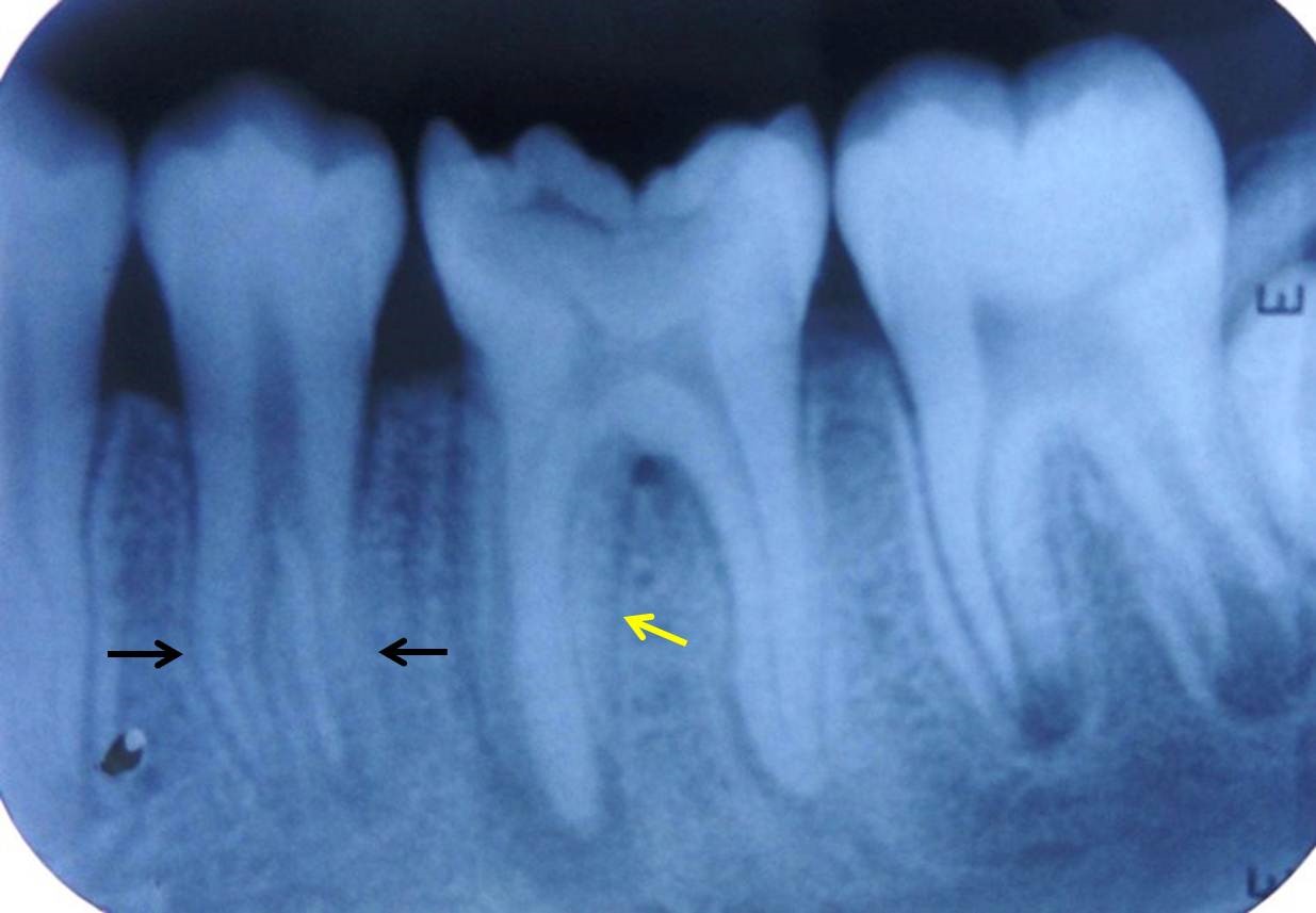
Figure 1: Intra oral periapical radiograph showing two roots in permanent mandibular right second molar (black arrows). Extra root (Radix Paramolaris) noticed in first molar (yellow arrow)
FIGURE 2
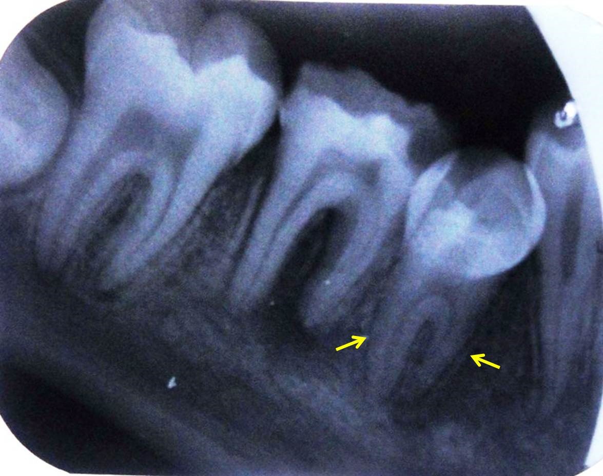
Figure 2: Evidence of two roots in permanent mandibular left second molar on radiograph (yellow arrows)
FIGURE 3
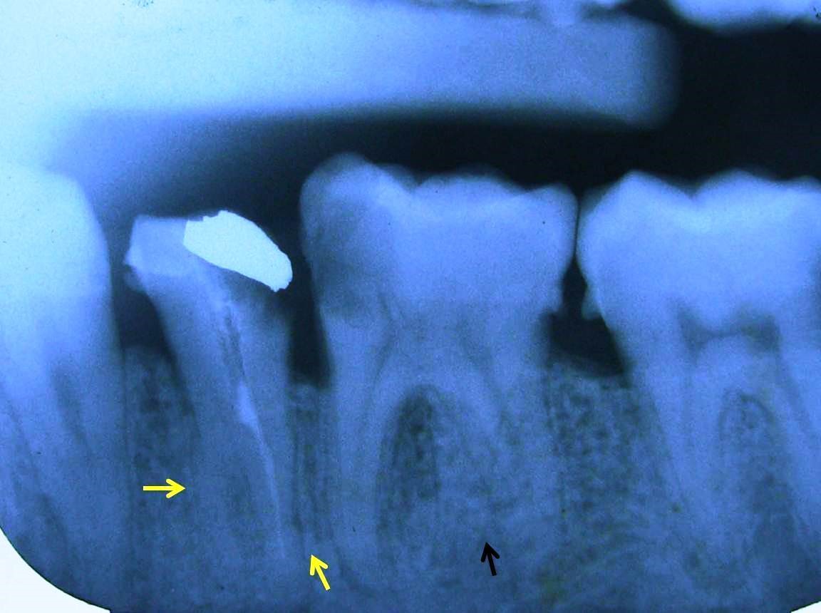
Figure 3: Radiograph showing presence of two roots (yellow arrows) in permanent mandibular right second molar. Radix Entomolaris was also present in first molar (black arrow)
Tables at a glance
Figures at a glance