Premolarization of the Permanent Maxillary Second Molars- Report of A Rare Case
Received Date: May 23, 2023 Accepted Date: June 23, 2023 Published Date: June 26, 2023
doi: 10.17303/jdoh.2023.10.105
Citation: Nagaveni NB (2023) Premolarization of the Permanent Maxillary Second Molars- Report of A Rare Case. J Dent Oral Health 10: 1-8
Abstract
Permanent maxillary second molars are usually seen with four cusps, with two buccal and two lingual cusps. Variation in size and shape rarely occur and attains both anthropological point of interest and even to dentists. Knowledge of occurrence of rudimentary or less number of cusps in any teeth is utmost important among clinicians. The present article describes occurrence of “premolarization” (mimicking of premolars) of permanent maxillary second molars which is not reported so far in the scientific literature. Therefore, the current case report is the pioneer case of a rare dental anomaly which alarms both anthropologists and dentist.
Keywords: Dental Anomaly; Maxillary Second Molar; Malformed Molars; Premolarization; Two Cusped Molars
Introduction
Permanent maxillary second molars are two in number, one right and the other one left situated posteriorly to first molars and anteriorly to third molars [1]. It supplements the first molar in function. In description of morphology of this tooth, direct comparisons can be made with the first molar including its form and development. Variation in tooth size and shape do exist and when they occur, show disturbances in the tooth formation stage. Dental anomalies associated with tooth size and shape call for an anthropological point of interest as well as to dentist [2].
Two types of permanent maxillary second molars exist based on the number of cusps (Figure 1). The first type is four cusped form and the second one is three cusped form. The first type is most frequently seen with two buccal and two lingual cusps [1]. The mesiolingual cusp is normally considerably wider than the distolingual cusp compared to maxillary first molars where the size of the lingual cups is closer to the same size. With respect to second type over one third of maxillary second molars are found with only three cusps which is popularly called by a term “tricuspid form.” In this form usually the distolingual cusp is missing,so it has just one large lingual cusp and two buccal cusps [1]. Very rarely the fifth cusp i.e., cusp of carabelli is found in these teeth. It is not uncommon to see more supplemental grooves as well as accidental grooves and pits on the occlusal surface of a maxillary second molar than are usually found on a maxillary first molar [1].
Presence of maxillary second molars has got clinical implications. Compared to first molars, maxillary second molars constitute second most teeth in the dental arch to provide proper occlusion. Second molars are also important for proper chewing and grinding as done by first molars. Therefore, clinical presence of second molars is very essential for the general overall well-being of the patient.
Sometimes maxillary second molars present extremely rare variation in both size and shape. Therefore, the purpose of this article is to present a case of maxillary second molar mimicking like maxillary premolar. Reduction in number of cusps and size is of great anthropologic interest as it shows changes in the evolution. Extensive literature on hominid evolution and dental anthropology shows no description of such rare dental anomaly of premolarization (shaped like premolar) of permanent maxillary second molars.
Case Report
An 18-year-old male patient reported to a private dental office seeking treatment for his irregular set of teeth. Patient was moderately built and well-nourished with no obvious features observed. On intraoral examination, patient exhibited complete permanent dentition with third molars still not erupted. When all the teeth were examined, the maxillary second molars appeared very small and almost looked like premolars (Figure 2 and 3). In mandibular arch all teeth were found in normal shape and size. Teeth were in normal occlusion with absence of any other dental anomalies.Impression of the maxillary dentition was taken and examination cast was made for further and detailed examination of all teeth (Figure 4). Permanent both right and left maxillary second molars had only two prominent cusps, one buccal and one lingual cusp with central groove separating the two cusps (Figure 2 and 3). The developmental grooves were found self-cleansing and non-retentive. The right second molar which looked like a premolar was rotated mesially. Measurement was carried out from mesial to distal and from buccal to lingual aspect using vernier caliper which showed the values almost similar to the values of maxillary premolars. Prominent fifth cusp called “cusp of carabelli” was noticed on both right and left permanent maxillary first molars (Figure 2 and 3). Finally based on clinical findings and literature search, this case was diagnosed as “Premolarized maxillary second molar” (i.e., maxillary second molar mimicking maxillary premolars). As developmental grooves were non retentive and self-cleansing regular proper tooth brushing was advised to the patient.
Discussion
Normally, two types of maxillary second molars are existing, when its detailed occlusal form is observed. From occlusal view, maxillary second molar exhibits rhomboidal shape which is most frequent encountered in all human beings. However, the acute angles of the rhomboid are less and the obtuse angles are greater when compared with the first molar. The buccolingual diameter of the crown is about equal, but the mesiodistal diameter is approximately 1 mm less [1]. Compared to first molar, the msiobuccal and mesiolingual cusps are quite large and well developed. But the distobuccal and distolingual cusps are smaller and less well developed. When a calibration made of the crown at the greatest diameter buccally and lingually of the distal portion is considerably less than one made at the greatest diameter buccally and lingually of the mesial portion, showing more convergence distally than the maxillary first molar. No fifth cusp is evident. The second type shows more resemblance to a typical third molar form. The distolingual cusp is poorly developed and makes the development of the other three cusps predominate. This result in a “heartshaped” or “tricuspid” form from the occlusal aspect that is more peculiar of the maxillary third molar [1].
The prevalence of variations in occlusal anatomy of teeth is reported more in maxilla compared to mandible [3]. Dental anomalies may be found localized to one tooth,or can be generalized to involving all the teeth. Sometimes they are reported as a part of syndromic or systemic disorders.
In 1922, World famous paleontologist Gregory described that among the major races of human beings the tooth crown morphology hardly varied dating back to early of the 20th century. However, there are numerous reports showing occurrence of extra cusps or roots, and variation in shape in humans [2-10], have identified and published various dental anomalies in Indian races. In 2015, authors described occurrence of permanent mandibular second premolar mimicking permanent mandibular second molar in an Indian patient which is too a rare phenomenon [4]. The authors coined the term “molarization” of premolar to this particular dental anomaly [4]. Formation of metaconulid or cusp 7 in permanent mandibular molars have been identified in humans [2]. Permanent maxillary third molar having multiple cusps (six supplemental cusps) in association with dens evaginatus is reported in 2013 [3]. In this case the maxillary third molar almost appeared like a flower. Reports on association of accessory roots like radix entomolaris and radix paramolaris which are also anthropological point of interest are seen in the literature [5-7]. Even in primary dentition extra cusp formation in molars too has been reported in the literature [8-10].
Human teeth are particularly used in anthropological research purpose as they have the advantage of observability,heritability, variability and preservability. Observability research using teeth involves skeleton, fossils and in the living. In heritability a strong genetic trait necessary for tooth development and expression of that trait in next generation is mainly studied. Variability is the one where measurement of traits is carried out which usually varies between populations and within a population. Finally, the preservability using teeth in anthropologic research is done for forensic records and experiments are made in fossils [2].
The present case report shows importance of further studies to be done in the future to evaluate its prevalence in different ethnic, races of the population around the globe. The limitation of this article is that presence of this type of variation was not examined in parents and other siblings in order to rule out the genetic influence on this dental rarity. However, the current report throws light on the existence of this type of dental variation in an Indian population thereby calling for the future studies and further research in the same population pertaining to premolarization of maxillary second molars.
- Ash MM, Nelson SJ (2003) Wheeler’s Dental Anatomy, Physiology and Occlusion. 8th ed. Noida, India: A Division of Reed Elsevier, Elsevier 165-70.
- Nagaveni NB (2008) Occurrence of cusp 7 (Metaconulid) in permanent lower first molars – Report of 4 cases and review of literature. International Journal of Dental Anthropology 13: 22-7.
- Nagaveni NB, Umashankar KV (2013) Maxillary molar with Dens evaginatus and multiple cusps – Report of a rare case and literature review. International Journal of Oral Health Sciences 3: 92-7.
- Nagaveni NB, Umashankar KV, Radhika NB, Mohan M (2015) Molarization of the mandibular second premolar in an Indian patient: Report of a rare case. Annals of Bioanthropology 3: 33-5.
- Nagaveni NB, Umashankar KV, Radhika NB (2012) A retrospective analysis of accessory roots in mandibular molars of Indian pediatric patients. International journal of Dental Anthropology 20: 38-46.
- Nagaveni NB, Umashankara KV (2009) Radix entomolaris in permanent mandibular first molars: Case reports and literature review. General Dentistry (General Discussion) 57:e24-9.
- Nagaveni NB, KV Umashankara (2012) Radix entomolaris and paramolaris in children – A review of the literature.Journal of Indian Society of Pedodontics and Preventive Dentistry 30: 94-102.
- Nandini DB, Nagaveni NB, Deepak BS, Poornima P (2019) Bilateral dens evaginatus in deciduous first molars: A rare finding. Journal of Medicine, Radiology, Pathology and Surgery 6: 9-11.
- NB Nagaveni, KV Umashankara, NB Radhika, RS Garewal (2009) “Paramolar Tubercle” in the primary dentition: Case reports and literature review. International Journal of Dental Anthropology 14: 12-8.
- Nagaveni NB, Umashankara KV, Poornima P, Subba Reddy VV (2014) Paramolar tubercle (Parastyle) in primary molars of Davangere (India) children: A retrospective study.International journal of oral health sciences 4: 18-22.
FIGURE 1
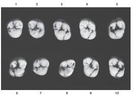
Figure 1: picture showing normal occlusal morphology of the permanent maxillary second molar. (Courtesy: Ash and Stanley [1])
FIGURE 2
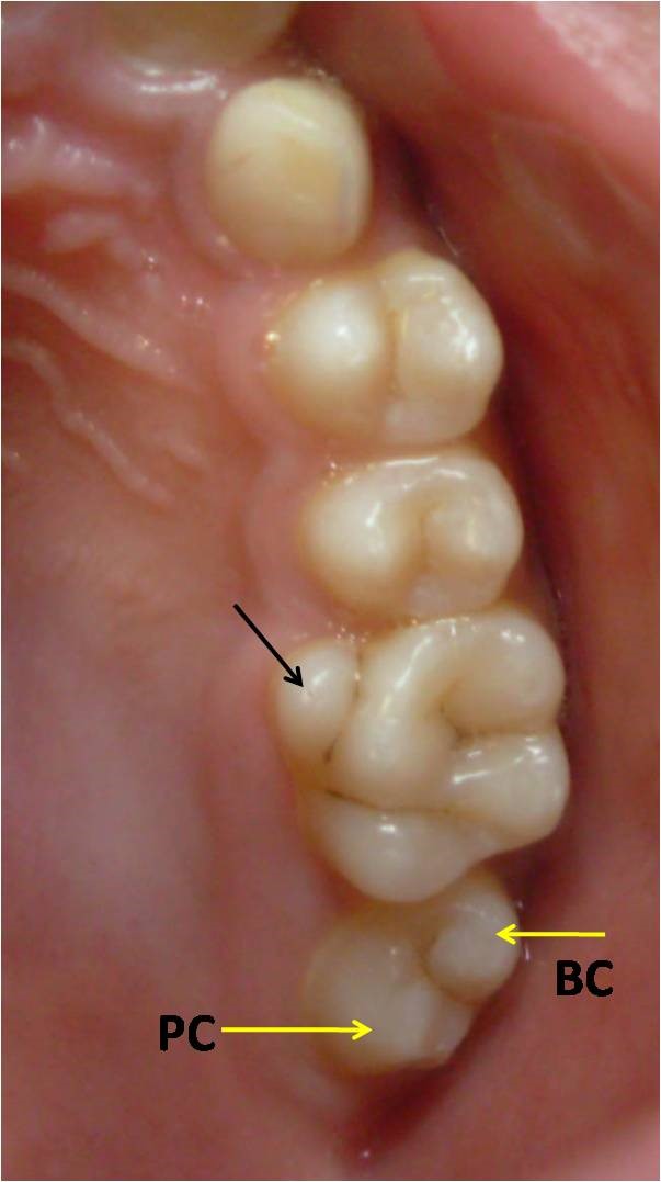
Figure 2: Maxillary left second molar mimicking a maxillary premolar with one buccal (BC) and one lingual cusp (LC). Prominent cusp of carabelli can be seen on first molar (black arrow)
FIGURE 3
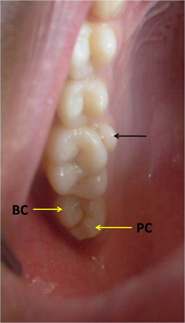
Figure 3: Premolarization (premolar like molar) of maxillary right second molar with two cusps (one buccal – BC and one lingual cusp - LC). Cusp of carabelli also noticed (black arrow)
FIGURE 4
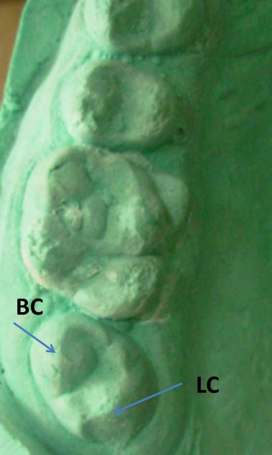
Figure 4: Dental cast showing premolar like maxillary second molar which is rotated (only two cusps, BC – buccal cusp and LC – lingual cusp)
Figures at a glance