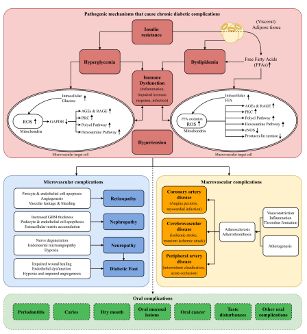
Figure 1: Schematic illustration of pathogenetic mechanisms that cause chronic diabetic complications
Figure 1: Schematic illustration of pathogenetic mechanisms that cause chronic diabetic complications
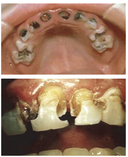
Figure 2 and 3: Early childhood caries and Rampant caries (We thank the authors for granting permission to use this figure)
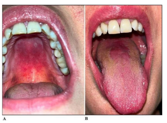
Figure 4: Clinical presentation of Candidiasis
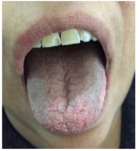
Figure 5: Clinical presentation of Xerostomia
Tables at a glance
Figures at a glance