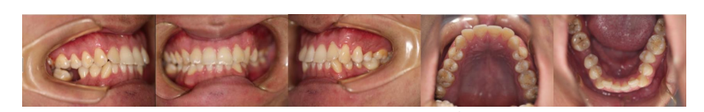
Figure 1: Pretreatment intraoral photographs showing intraoral plaque and soft tartar build-up, red and swollen gums and missing right lower jaw first molar
Figure 1: Pretreatment intraoral photographs showing intraoral plaque and soft tartar build-up, red and swollen gums and missing right lower jaw first molar
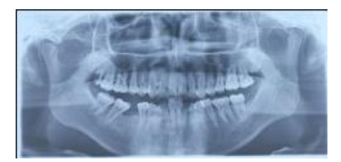
Figure 2: Pretreatment panoramic radiograph showing a severe inclination of the lower right second molar and third molar proximally
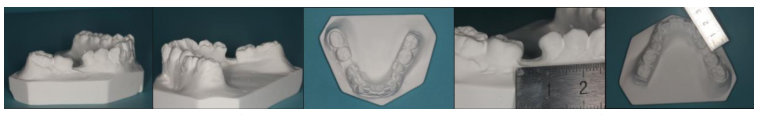
Figure 3: Pretreatment measurement of the patient's intraoral plaster model showing a lack of adequate restoration space
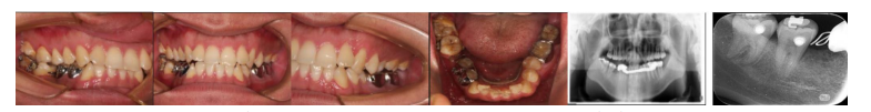
Figure 4: Insertion of a temporary anchorage devices and bonding of a fixed cast ring aligner
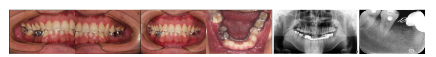
Figure 5: The implant gap gradually increased after about two months of orthodontic treatment
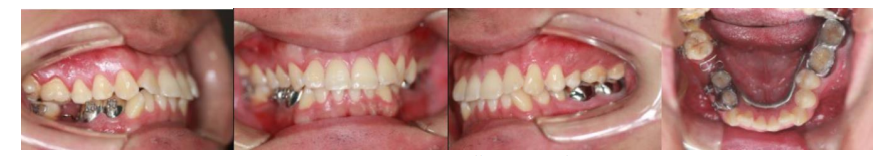
Figure 6: Intraoral photographs showing sufficient space for implant restoration
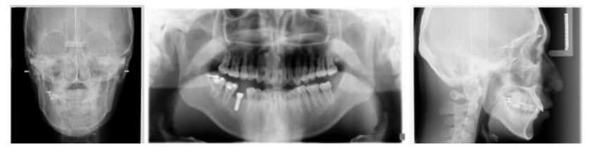
Figure 7: Radiographs showing implant placed in the right mandibular first molar region
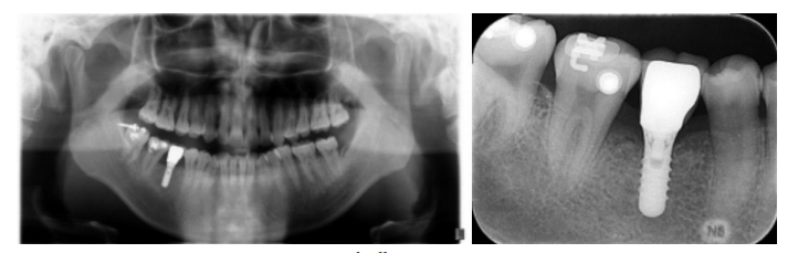
Figure 8: Patients with all-ceramic crown restorations
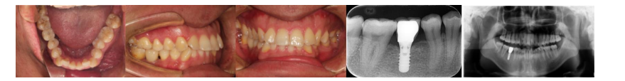
Figure 9: Intraoral photographs and radiographs showed that the implant denture was functioning well in the mouth
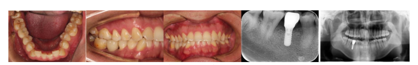
Figure 10: One year and three months after the end of the patient's multidisciplinary combined treatment
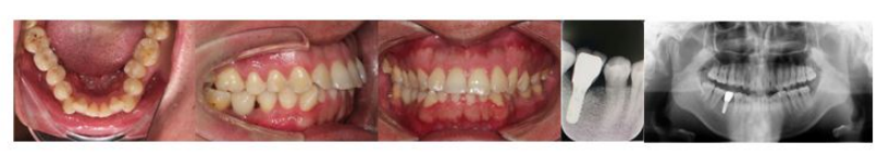
Figure 11: Three year and five months after the end of the patient's multidisciplinary combined treatment
Figures at a glance