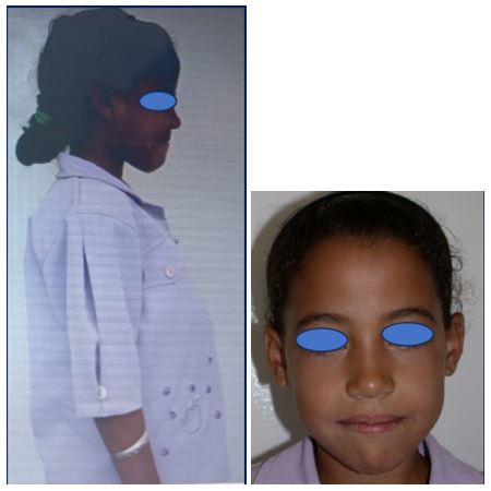
Figure 1: Exobuccal photos showing increased abdominal perimeter caused by the hepatosplenomegaly, and ‘’asian facies’’ appearance
Figure 1: Exobuccal photos showing increased abdominal perimeter caused by the hepatosplenomegaly, and ‘’asian facies’’ appearance
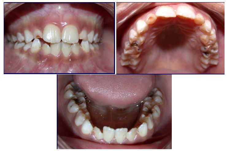
Figure 2: Pictures of the oral cavity
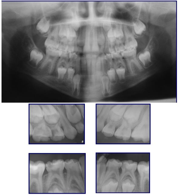
Figure 3: Panoramic X-ray complemented by four retro-alveolar X-rays showing dentino-pulpal damage on tooth 54 and 85, dentinal caries on tooth 65 and 75
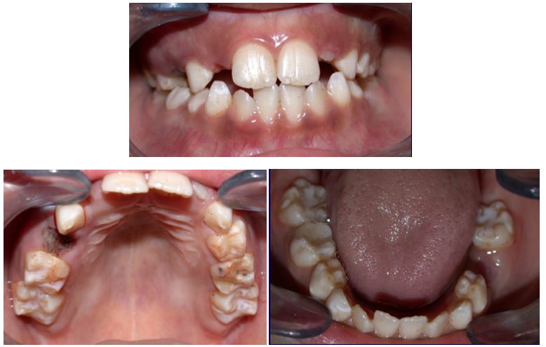
Figure 4: Final result after extracting tooth 54 and 75, doing restorations on tooth 55 and 85, and applying sealants on the four first permanent molars
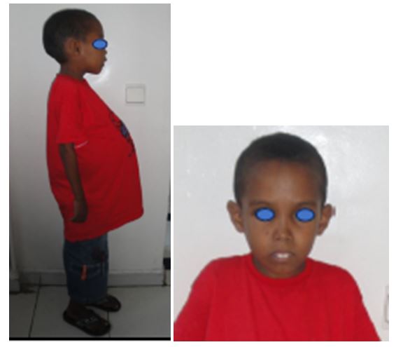
Figure 5: Photos showing increased abdominal perimeter caused by the hepatosplenomegaly, and ‘’asian facies’’ appearance
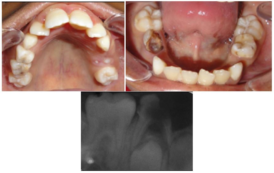
Figure 6: Photos of the oral cavity showing the parulic abscess related to damaged tooth 85, and the retro-alveolar X-ray taken for tooth 85 showing dentino-pulpal damage with inter-radicular radiolucency.
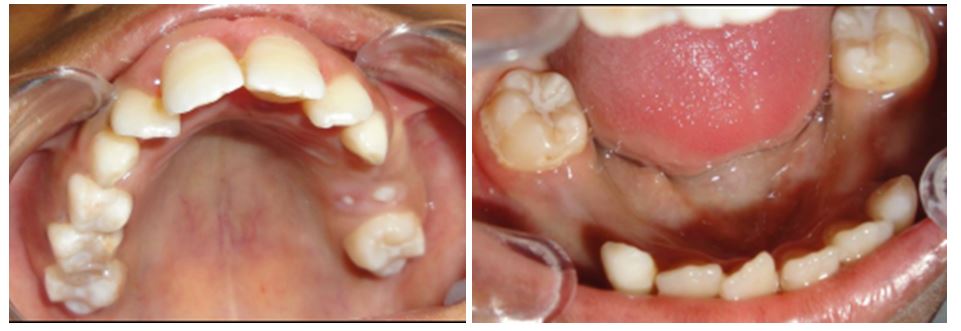
Figure 7: Final result after extraction tooth 85 and applying sealants for the four first permanent molars
Figures at a glance