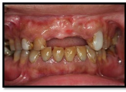
Figure 1 Preoperative labial view
Figure 1 Preoperative labial view
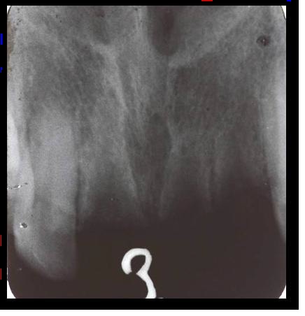
Figure 2 Preoperative peri-apical radiograph
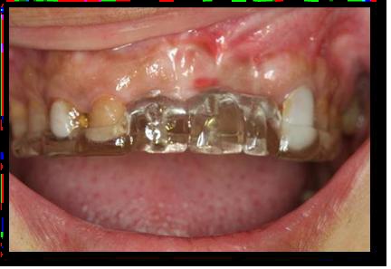
Figure 3 Surgical stent in position
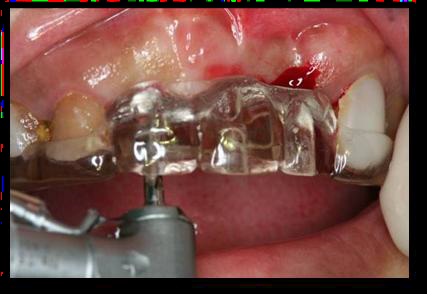
Figure 4 Small, initial penetration through the mucosa and into bone.
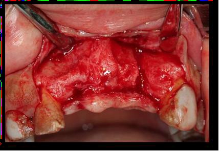
Figure 5 Reflection of full muco-periostal flap
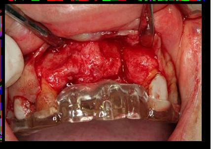
Figure 6Surgical stent in position after flap reflection (supported by adjacent teeth).
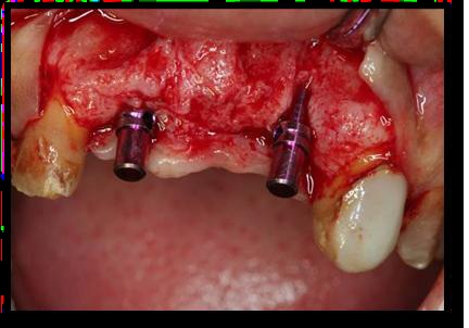
Figure 7 Guide Pin in osteomy prepared site (check for parallelism and angulations)
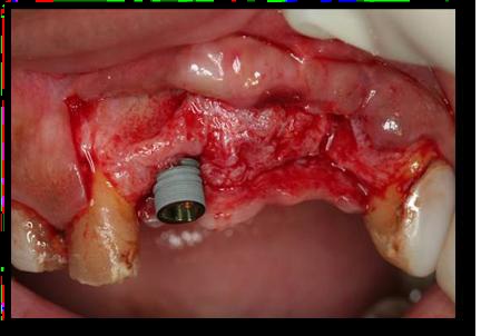
Figure 8 Placement of implant fixture
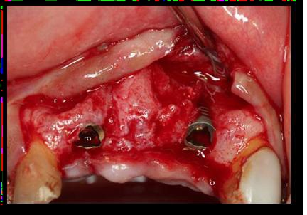
Figure 9 Both Implant fixtures in position
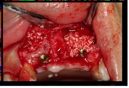
Figure 10 Inlay guided bone regeneration
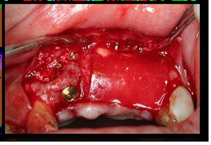
Figure 11 GBR membrane in position
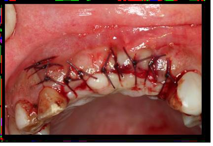
Figure 12 Flap Closer (interrupted suture)
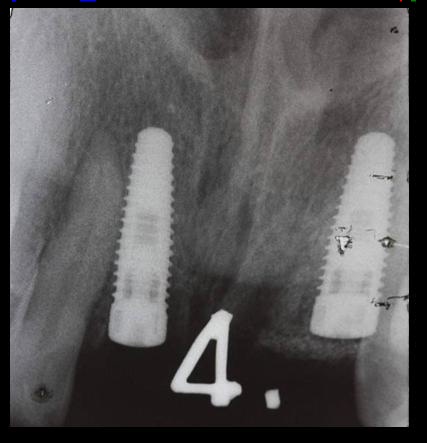
Figure 13 Peri-apical X-ray after first surgery
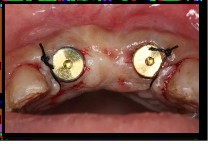
Figure 14 Healing abutment after second surgery
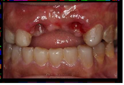
Figure 15 Gingival margin healing
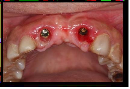
Figure 16Occlusal view of gingival margin healing
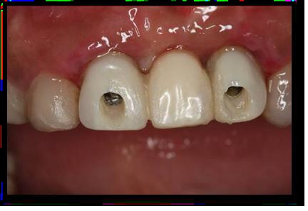
Figure 17 Screw Retained temporary bridge
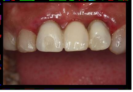
Figure 18 Closing access opening for screw retained temporary bridge (composite resin)
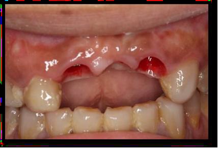
Figure 19 Gingival margin before final restoration
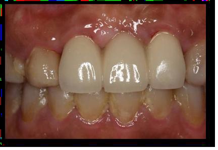
Figure 20 Labial view of the final restoration
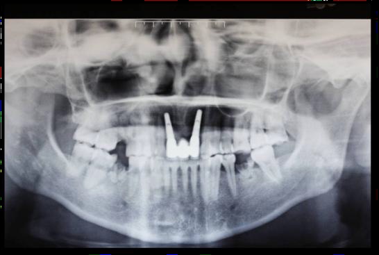
Figure 21 Panoramic X-ray of final treatment