
Figure 1 Angiofibromas presenting in a butterfly
Figure 1 Angiofibromas presenting in a butterfly
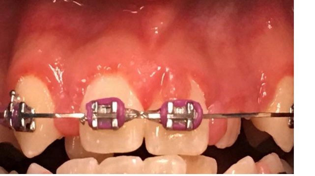
Figure 2 Gingival enlargement surrounding maxillary
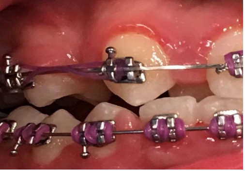
Figure 3 Gingival enlargement surround maxillary
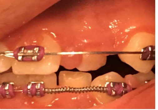
Figure 4 Extensive gingival overgrowth around maxillary
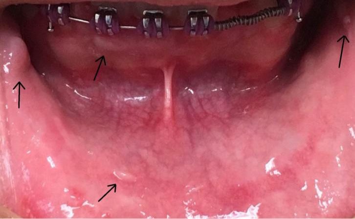
Figure 5 Inraoral fibromas on labial mucosa andfibroticplaqueson gingiva and lower lip (arrows)
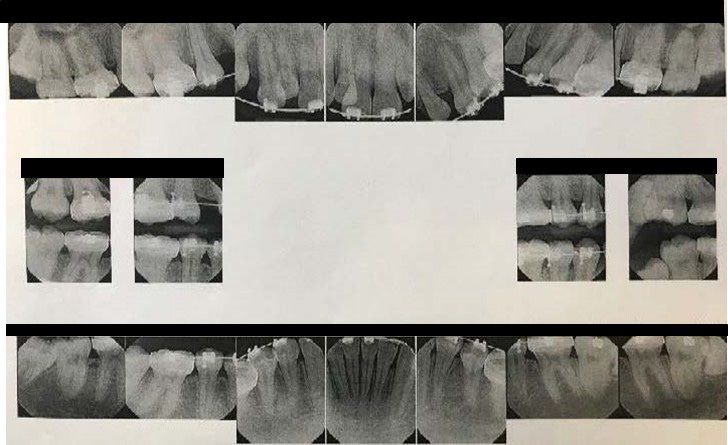
Figure 6 FMX at the time of the diagnostic clinic. No pathological anomalies detected
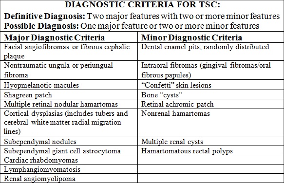
Table 1 Major and Minor Diagnostic Criteria for TSC (12,13)