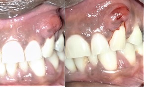
Figure 1 A). Preoperative image showing swelling and pus discharge in relation to 23, B). Incision and drainage done.
Figure 1 A). Preoperative image showing swelling and pus discharge in relation to 23, B). Incision and drainage done.
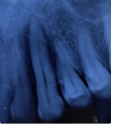
Figure 2 Preoperative Radiograph
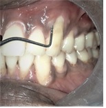
Figure 3 1-week post-operative image showing a probing depth of 8mm
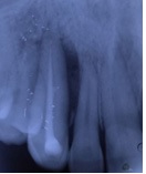
Figure 4
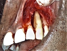
Figure 5 Full-thickness mucoperiosteal flap raised showing the osseous defect
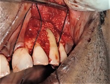
Figure 6 Osseous defect filled with the bone graft (allograft)
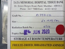
Figure 7 Freeze – Dried, Irradiated Amnion Membrane
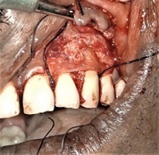
Figure 8 Amniotic membrane placed over the bone graft in osseous defect
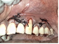
Figure 9 Flap approximated with interrupted sutures
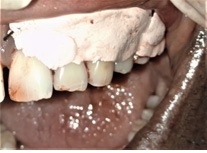
Figure 10 Coe-Pak dressing placed over the surgical site
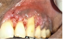
Figure 11 1-week Post-operative Healing
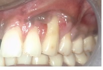
Figure 12 1-month Post-operative Healing
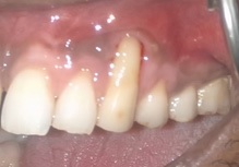
Figure 13 6 Months Post-operative Healing
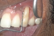
Figure 14 Reduction of Probing depth to 3mm
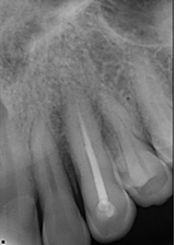
Figure 15 6 Months postoperative radiograph showing bone fill in surgical site